Clinical Image, J Otol Rhinol Vol: 8 Issue: 3
Unusual Ear Mass
Anita Jeyakumar1* and Matthew Keisling2
1Department of Otolaryngology, Akron Children’s Hospital 215 W Bowery St, Ste 3210, Akron, Ohio, USA
2Department of Pathology, Akron Children’s Hospital, Akron, Ohio, USA
*Corresponding Author : Anita Jeyakumar Department of Otolaryngology, Akron Children’s Hospital 215 W Bowery St, Ste 3210, Akron, OH 44308, USA Tel: 330-543-4930 Fax: 330-543-4931 E-mail: ajeyakumar@akronchildrens.org
Received: September 18, 2019 Accepted: September 25, 2019 Published: October 01, 2019
Citation: Jeyakumar A, Keisling M (2019) Unusual Ear Mass. J Otol Rhinol 8:3. doi: 10.4172/2324-8785.1000376
Abstract
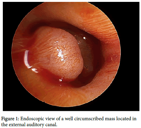
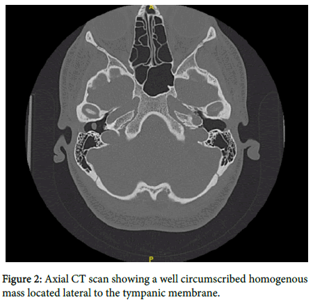
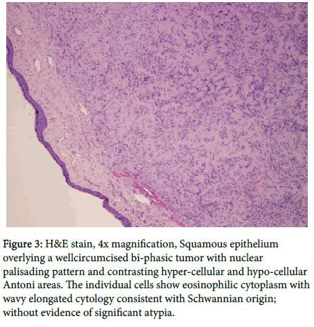
17 year old female with a history of stage IVS neuroblastoma diagnosed at the age of 4 months. She received cis-retinoic acid post radiation and completed all therapy at the age of 4 years. During a routine audio logic visit, she was noted to have a mass in her right ear (Figure 1). A CT scan was done which showed a small homogenous well circumscribed mass in the left external auditory canal (Figure 2). The mass was biopsied in both left and right ear and the pathology is shown in Figure 3, with a diagnosis of a Schwannoma.
Keywords: Tympanic membrane; Neuroblastoma
About the Study
17 year old female with a history of stage IVS neuroblastoma diagnosed at the age of 4 months. She received cis-retinoic acid post radiation and completed all therapy at the age of 4 years.
During a routine audio logic visit, she was noted to have a mass in her right ear (Figure 1). A CT scan was done which showed a small homogenous well circumscribed mass in the left external auditory canal (Figure 2). The mass was biopsied in both left and right ear and the pathology is shown in Figure 3, with a diagnosis of a Schwannoma.
Figure 3: H&E stain, 4x magnification, Squamous epithelium overlying a wellcircumcised bi-phasic tumor with nuclear palisading pattern and contrasting hyper-cellular and hypo-cellular Antoni areas. The individual cells show eosinophilic cytoplasm with wavy elongated cytology consistent with Schwannian origin; without evidence of significant atypia.
Schwannomas are slow-growing benign tumors. In the head and neck region, Schwannomas most commonly appear as acoustic neuroma (25–45%) [1]. Schwannomas are rarely diagnosed in the external auditory meatus but there have been a few reports in the literature [2-6]. The differential diagnosis includes other soft tissue neoplasms: fibroma, chondroma, and leiomyoma. The mass was treated by complete surgical excision.
References
- Colreavy MP, Lacy PD, Hughes J, Bouchier-Hayes D, Brennan P, et al. (2000) Head and neck schwannomas-a 10 year review. J Laryngol Otol 114: 119-124.
- Galli J, D'ecclesia A, La Rocca LM, Almadori G (2001) Giant schwannoma of external auditory canal: A case report. Otolaryngol Head Neck Surg 124: 473-474.
- Gross M, Maly A, Eliashar R, Attal P (2005) Schwannoma of the external auditory canal. Auris Nasus Larynx 32: 77-79.
- Lewis WB, Mattuchi KF, Smilari T (1995) Schwannoma of the external auditory canal: an unusual finding. Int Surg 80: 287-290.
- Harcourt JP, Tungekar MF (1995) Schwannoma of the external auditory canal. J Laryngol Otol 109: 1016-1018.
- Topal O, Erbek SS, Erbek S (2007) Schwannoma of the external auditory canal: a case report. Head Face Med 3: 6.
 Spanish
  Chinese
  Russian
  German
  French
  Japanese
  Portuguese
  Hindi
Hindi 


