 Spanish
  Chinese
  Russian
  German
  French
  Japanese
  Portuguese
  Hindi
Hindi Review Article, Dent Health Curr Res Vol: 4 Issue: 2
History of Digital Detectors in Intraoral Radiography
Midori Yoshida*, Hozumi Yoshihara and Eiichi Honda
Department of Oral and Maxillofacial Radiology, Tokushima University, Graduate School, Japan
*Corresponding Author : Midori Yoshida
Department of Oral and Maxillofacial Radiology, Tokushima University, Graduate School, Japan
Tel: +81-88-633-5361
E-mail: midori2@tokushima-u.ac.jp
Received: April 11, 2017 Accepted: May 08, 2018 Published: May 15, 2018
Citation: Yoshida M, Yoshihara H, Honda E (2018) History of Digital Detectors in Intraoral Radiography. Dent Health Curr Res 4:2. doi: 10.4172/2470-0886.1000135
Abstract
The first digital sensor used in intraoral radiography, RadioVisioGraphy was introduced in 1987. It was based on the activity of a charged-Coupled Device (CCD) sensor. Later on, other digital sensors became commercially available, including the Digora featuring an imaging plate coated with photostimulable phosphor (PSP) in 1994 and the complementary metal-oxide semiconductorbased sensor, CDR active pixel sensor, in 1998. These technologies have recently undergone considerable improvement, and several dental clinics have switched from film-based systems to digital imaging. In the early days, digital sensors were considered inferior to film in clinical diagnostics, but according to recent research results, there is no longer any significant difference because of the improvement in performance. However, our latest research using a new evaluation method and a precise phantom model indicated some differences between the performance of CCD and PSP systems and further differences between PSP systems of different function and performance.
Keywords: Digital; Intraoral radiography; PSP; CCD
Introduction
In dentistry, radiography is one of the indispensable examinations. The two commonly used methods are intraoral and panoramic radiography. Lately, a computed tomography technique developed for use in dentistry, the so-called cone beam computed tomography has been widely used as well. Although silver halide emulsion-coated films are still in use in intraoral and panoramic radiography, radiography using digital detectors is now becoming mainstream. Until the advent of intraoral digital sensors, the development of film-based technology consisted mainly of the improvement in sensitivity and exam speed (F-speed film being the fastest one). However, many famous film manufactures, such as Fuji Film (Kanagawa, Japan), Agfa (Mortsel, Belgium), and Eastman Kodak (Rochester, NY) are now withdrawing from the production of intraoral films because of the spread of digital detectors. The main cause is that since the computed radiography (CR) system developed by Fuji Film has replaced the screen-film system, the demand for film in medicine has reduced dramatically [1]. CR technology uses a photostimulable phosphor (PSP) imaging plate instead of film. Currently, the most significant manufacturer of intraoral films is Carestream Health (Rochester, NY), part of the Onex (Toronto, Canada) family of companies, and successor of Eastman Kodak in dental imaging technology production since they sold Kodak Health Group to Onex Corporation [2]. Carestream Health manufactures Ultra-speed and Insight films for intraoral radiography even today.
Meanwhile, the improvement of imaging sensors developed for general use has been tremendous. The first one, the charged-coupled device (CCD) sensor was inserted into a digital camera by Kodak [3]. Subsequently, color CCD was used in a personal video camera, and a color complementary metal-oxide semiconductor (CMOS) sensor was also developed later on. Technological features such as small size, high resolution, and power saving have advanced gradually, because CCD and CMOS sensors have become widely used in personal digital cameras including mobile phones. These sensors have been used in intraoral radiography before.
Sensors for intraoral digital radiography
Many types of detectors for radiography are used in dentistry, such as CCD, CMOS, flat panel detector, and PSP imaging plate. Of these, the CCD image sensor, the CMOS image sensor, and the PSP imaging plate are used in intraoral radiography.
Intraoral digital radiography has the following characteristics compared with film radiography:
Advantages
• Immediate image display after exposure (solid-state type)
• Less patient exposure
• Image processing available
• No age-related deterioration
• No chemicals, such as developer and fixer
• Less storage space
• Image data transfer is possible through the internet
• Wide dynamic range (PSP type)
Disadvantages
• Smaller sensor size (solid-state type)
• No flexibility (solid-state type)
• Expensive
• Narrow dynamic range (solid-state type)
• Difficulty of infection control for reuse
• Low spatial resolution (PSP type)
The CCD image sensor was invented at Boyle and Smith at AT&T Bell Laboratories in 1970 [4]. CCD can directly absorb electromagnetic rays such as visible light, infrared rays, ultraviolet rays, and low-energy X-rays and transduce them into charge, which is electrical energy. However, the wavelength band of 20 to 70 keV used in intraoral radiography is shorter than the sensitive wavelength band. Therefore, general CCD image sensors are not used alone in intraoral radiography. It is necessary to convert the wavelength to increase sensitivity, usually by a scintillator. In many cases, a CsI (Tl) scintillator with an emission peak of around 550 nm (green) is used, which converts X-ray into light. Moreover, the CCD sensor is coupled to a fiber optic plate (FOP) with a scintillator (Figure 1). The structure is the same in a CMOS sensor. Currently used CCD area image sensors are classified into five types based on the charge transfer method: frame transfer (FT), full-frame transfer (FFT), interline transfer (IT), frame interline transfer (FIT), and linear image sensor (one-dimensional). The FFT type is used in the X-ray detector. CCD sensors have also been used in dentistry [5].

Figure 1: Sectional structure of representative solid-state image sensor.
The CMOS sensor is similar to the CCD sensor. The transfer method is decisively different from that of the CCD sensor. In the CCD sensor, the charge formed by X-ray is temporarily stored in the potential well in each pixel, and the charge is transferred to the adjacent well by using sequentially shifted voltage (Figure 2 and Figure 3). However, in a CMOS sensor, each pixel is directly connected to the output through each independent amplifier (Figure 4). Charges formed at each pixel are immediately transmitted to the output. Although it is not a concern in intraoral radiography, it is known that there is a risk of distortion when shooting high-speed movies due to the time difference between the first and the last transmitted charges. Metal-oxide semiconductor (MOS) type image sensors are divided into two types: passive pixel sensors (PPSs) and active pixel sensors (APSs). The PPS does not have the function of amplifying the signal charge in each pixel, whereas the APS is capable of this. APS sensors are used for intraoral radiography. APS technology was first presented [6]. A color sensor requires three types of elements corresponding to red, green, and blue (RGB). On the other hand, CMOS handles the RGB signal using one element. A currently used CMOS image sensor for intraoral radiography is the APS type, and it is used together with a CsI (Tl) scintillator or a phosphor screen, similarly to the CCD image sensor. PSPs used in intraoral radiography are the same as PSP imaging plates manufactured by Fuji Film described above. A PSP BaFBr:Eu (Eu is doping) is coated on a thin polyethylene terephthalate plate. Fuji Film found BaFBr:Eu optimal among more than 1000 fluorescent substances. In the crystal, ionization occurs by X-ray energy, and the ionizing electron is trapped in BaFBr. The crystal emits fluorescence by releasing the electron upon laser irradiation. An image is created by measuring the amount of fluorescence because it is proportional to the exposure dose (Figure 5).

Figure 2: Structure of full frame transfer (FFT) type CCD image sensor. Arrows show charge flow. The charge moves sequentially from a register to the adjacent register. After charges reach the end of a vertical shift register, the charges move to the horizontal shift register.

Figure 3: Charge transfer of CCD image sensor.
(a) Electric charge is formed by X-ray exposure. The charge is temporarily stored in each vertical shift register.
(b) Each charge moves to the adjacent register by applying a voltage. The charge reaching the end of a vertical shift register moves to a horizontal shift register. (c) and (d) Each charge moves sequentially.

Figure 4: Structure of metal-oxide semiconductor (MOS) image sensor.
Each photodiode corresponding to each pixel has independent amplifier and pixel selection switch. Because it is impossible to transmit all charges in each pixel simultaneously to the output, A time difference arises between the first and the last transmitted charge. The difference produces motion artifact of an image.

Figure 5: Principle of photostimulable phosphor (PSP) system.
(a) The crystal of BaFBr:Eu is exposed.
(b) Ionization of Eu occurs, and Eu2+ turns into Eu3+. The ionizing electron is trapped in BaFBr.
(c) In reading process, the exposed crystal is irradiated by laser.
(d) The laser irradiation affects the electron trap, and the electron recombines with Eu3+. Therefore Eu3+ returns to Eu2+, and the energy difference produces fluorescence. The fluorescence is measured by detector.
Comparison of CCD and CMOS
The image quality of a CCD image sensor is superior to that of a CMOS image sensor in low signal intensity, because dark current noise is low. However, the current production volume of CMOS is much higher than that of CCD for the advantages explained below. Today, image sensor production/utilization companies like Nikon (Tokyo, Japan), Canon (Tokyo, Japan), and Sony (Tokyo, Japan) have replaced CCD with CMOS. Moreover, Sony has discontinued the manufacturing of CCD products in 2015.
Advantages
• High-speed data transfer (frame rate) because each pixel is independently connected to the output
• Less expensive, because it can be made into one chip
• Low power consumption, because high voltage circuit is not necessary
• No blooming (#1) and smear (#2), because each pixel has an independent amplifier
• Wide dynamic range, because the saturation charge of each pixel can be controlled by the amplifier
Streaks above and below the bright spot in the image caused by bleeding electron charge from an overexposed pixel into other nearby pixel
Very bright spots in the image caused by stray electrons generated under the photodiode area that diffused into the vertical shift registers
Disadvantages
• Generation of fixed pattern noise, because a fixed amplifier is assigned for each pixel
• Low sensitivity
• Low homogeneity
• Low linearity
Early days of intraoral digital radiography
The first intraoral digital sensor was introduced in 1987 [7]. The system was called RadioVisioGraphy (RVG) (Trophy Radiologic, Vincennes, France), and it consisted of a CCD with an intensifying screen. It became evident that the spatial resolution was slightly inferior to that of film, but patient exposure was considerably reduced [8]. In clinical practice, no differences were found in the length of the root canal as pictured by RVG and conventional film images [9]. However, during the measurement of #15 Headstrom file, the length of non-enhanced RVG image was found to be shorter than that of conventional film image [10]. The new RVG type was presented in 1991 [11]. Its resolution was reported to be 11 Lp/mm compared with that of film, which was 14 line pairs/mm. The RVG system was considered to be particularly useful for endodontic treatment, because the image was immediately displayed after exposure. Another reason was that the exposure dose reduction rate was about 80% compared with D-speed film [12].
After the introduction of RVG, other direct digital intraoral sensors based on CCD were developed. Sens-A-Ray (Regam Medical System AB, Sundsvall, Sweden) was presented in 1992 [13]. The resolution was the same as that of RVG, and the sensitivity was over three times that of E-speed. However, the successful identification of holes in aluminum foil was reduced compared with D-speed and E-speed films [14].
VIXA/Visualix (Gendex Dental Systems, Monza, Italy) (VIXA in North America) and Flash Dent (Villa Sistemi Medicali, Buccinasco, Italy) were also developed for intraoral radiography, and those with RVG and Sens-A-Ray were compared for the determination of root canal length using a #10 file. All four sensors were proven unacceptably inferior to E-speed film [15].
The fourth-generation RVG, RVG32000 (Trophy Radiologic), was presented in 1993 [16]. The further-improved RVG-S (Trophy Radiologic) was introduced in 1995 [17]. It was reported to be superior to E-speed film in sensitivity for the detection of accessory/ lateral canals with zoom. The performance of CCD sensors is now considered as equivalent to film.
The PSP system was released in 1983 in medicine, but its introduction to dentistry happened quite late. The first PSP-based intraoral system was Digora (Soredex, Helsinki, Finland), which became commercially available in 1994 [18]. The next PSP system, DenOptix (Gendex Dental Systems) was presented in 1997 [19]. In early research, the performance in the in vitro detection of caries was reported to be similar between CCD and PSP systems [20]. Later on, it was reported that solid-state systems had better resolving power because of the higher contrast and smaller pixel sizes compared with PSP systems [21]. However, the PSP system was shown to produce higher image quality over a much wider exposure range than the CCD sensor [22]. Moreover, Digora was reported to have the best signal-to-noise ratio, and DenOptix was the worst among Visualix, CDR (Schick Technologies, Long Island City, NY), CDR-APS (Schick Technologies), and Digora [19].
Another solid-state sensor, the CMOS sensor was introduced considerably later than the CCD sensor in 1998 [19]. The first product was CDR-APS. No significant difference was reported between the performance of CCD and CMOS sensors in the detection of artificial periapical lesions [23]. In 2001, it was also reported that the detection ability of proximal caries was similar among CMOS, E-speed, and F-speed films [24].
Improvement of intraoral digital radiography
The performance of intraoral digital sensors such as the solidstate sensor and the PSP imaging plate has advanced rapidly. Spatial resolution was clearly inferior to that of analog film in the early days. Spatial resolution is closely related to pixel size. In the PSP system, the scanning interval also affects the spatial resolution. Although scanning interval can be made as short as possible, resolution does not improve when it is smaller than the aperture size of the scanner. Theoretically, the maximum resolution defined by Lp/mm is 1/(twice pixel size). Around 2000, the representative pixel size of the CCD sensor was 39 μm for Compuray (Yoshida, Tokyo, Japan), 45 μm for Sens-A-Ray, 48 μm for CDR, and 63 μm for VIXA, whereas that of the PSP was 42 μm for DenOptix and 70 μm for Digora [25]. Thus, the theoretical maximum resolution is 7.9 to 12.8 Lp/mm for CCD and 7.1 to 11.9 Lp/mm for PSP. The value was inferior to these of film. In another publication, it was reported that the pixel size of CMOS sensor (CDR-APS) was 40 μm [19].
The performance of the solid-state sensor has gone through tremendous improvement, because the sensor is being used in daily life in personal digital cameras. The improvement in spatial resolution is especially dramatic. In the 2005 report, the measured spatial resolution of a CCD sensor, RVG6000 (Eastman Kodak, Rochester, NY), a CMOS sensor, RVG-ui (Eastman Kodak), and the F-type speed film Insight (Eastman Kodak) were over 20 Lp/mm [26]. The pixel size of RVG6000 and RVG-ui was 18.5 and 19.5 μm. The spatial resolution of a currently used CMOS sensor, HDI-S (Rayance, Gyeonggi-do, Korea) reaches 33.7 Lp/mm [27].
Topics on recent intraoral digital sensors
Recent studies have shown no apparent clinical difference among intraoral detectors. The diagnostic accuracy of CCD, Dixi3 (Planmeca, Helsinki, Finland) and PSP, Digora PCT (Soredex) was reported to that of film systems (Ektaspeed, Eastman Kodak) in the detection of non-cavitated proximal caries and in measuring root canal working length [28,29]. Concerning the detection or root resorption, no significant difference has been reported among CCD (Sopro Imaging, Ac‑t Croup, La Ciotat, France), PSP (Digora optime, Soredex, Tuusula, Finland), and Ektaspeed film systems [30]. Similarly, no significant differences among CCD (Dixi3), PSP (Digora), and film (Ektaspeed) systems were observed [31].
However, our recent research showed a clear difference among different intraoral digital sensors [32]. A CCD sensor (Megadixel, Morita, Tokyo, Japan) and two types of PSP (Digora optime) (VistaScan perio, Durr dental AG, Bietigheinm-Bissingen, Germany) were used in the research. For comparison, a precise aluminum step wedge phantom with an accuracy of 10 μm was custom made, and a new evaluation method was devised. One evaluation value was contrast-to-noise ratio, which represents low contrast between thin objects such as minute bone regeneration during periodontal treatment. The other evaluation was low contrast value, which represents low contrast between thick objects such as molar proximal caries. These were defined by the following equation:
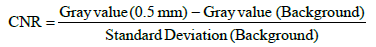
LCV Gray value(11mm) Gray value(10 mm)
where the Gray value (x mm) is the average for the aluminum step image at x mm (Figure 6).

Figure 6: Contrast noise ratio (CNR) and Low contrast value (LVC).
Digora: Digora Optime (PSP), VistaScan: VistaScan perio (PSP), Megadixel (CCD).
(a) CNR. Clinical standard exposure time is 1.6s for PSP and 0.8s for CCD for anterior teeth.
(b) LCV. Clinical standard exposure time is 3.2s for PSP and 1.6s for CCD for molars.
The following conclusions were obtained from the research: the CCD sensor system was indicated to be optimal for all diagnostic purposes, regardless of whether it could be clinically used. When the PSP system was selected, PSP systems having high bit depth like VistaScan perio were appropriate for diagnosing a slight bony change, and the PSP system having automatic exposure compensation function similar to that of Digora Optime was appropriate for diagnosing proximal caries of the molar. Thus, it is very important to select an appropriate sensor for diagnostic purpose, because the performance of digital sensors may influence the diagnosis.
Thus, our research implies that more rigorous experimenting is needed to clarify the difference in the performance of digital sensors, and the result will affect the diagnosis in more detail.
Acknowledgements
This study was supported by JSPS KAKENHI Grant Number JP16K1150800.
References
- Sonoda M, Takano M, Miyahara J, Kato H (1983) Computed radiography utilizing scanning laser stimulated luminescence. Radiology 148: 833-838
- Carestream (2015) 75 years of commitment. Carestream brochure. www.eurotehnika.hr/wp-content/uploads/2016/05/75-godina-predanosti-Kodak-Carestream.pdf
- DeLuce M Improved light sensitivity in color CCD image sensors. Kodak brochure. spectronet.de/story_docs/vortraege_2009/091103_vision/091105_0930_deluca_kodak.pdf
- Boyle WS, Smith GE (1970) Charge Coupled Semiconductor Devices. Bell Syst Tech J 49: 587-559
- Hamamatu Photonics. Image sensors. chapter 05. https://www.hamamatsu.com/resources/pdf/ssd/e05_handbook_image_sensors.pdf (accessed in April 2018)
- Schuster MA, Strull G (1966) A monolithic mosaic of photon sensors for solid-state imaging applications. IEEE Trans Electron Devices 13: 907-912
- Duret F, Coste BC, Duret, B (1988) The Radio Visiography (RVG): where reality surpasses radiological fiction a hope that is becoming reality. J Dent Pract Admin 5: 138-140
- Mouyen F, Benz C, Sonnabend E, Lodter JP (1989) Presentation and physical evaluation of RadioVisio Graphy. Oral Surg Oral Med Oral Pathol 68: 238-242
- Shearer AC, Horner K, Wilson NH (1990) Radiovisiography for imaging root canals: an in vitro comparison with conventional radiography. Quintessence Int 21: 789-794
- Shearer AC, Horner K, Wilson NH (1991) Radiovisiography for length estimation in root canal treatment: an in-vitro comparison with conventional radiography. Int Endod J 24: 233-239
- Benz C, Mouyen F (1991) Evaluation of the new RadioVisioGraphy system image quality. Oral Surg Oral Med Oral Pathol 2: 627-631
- Sanderink GC (1993) Imaging: new versus traditional technological aids. Int Dent J 43: 335-342
- Nelvig P, Wing K, Welander U (1992) Sens-A-Ray. A new system for direct digital intraoral radiography. Oral Surg Oral Med Oral Pathol 74: 818-823
- McDonnell D, Price C (1993) An evaluation of the Sens-A-Ray digital dental imaging system. Dentomaxillofac Radiol 22: 121-126
- Sanderink GC, Huiskens R, Stelt PF, Welander US, Stheeman SE (1994) Image quality of direct digital intraoral x-ray sensors in assessing root canal length. The RadioVisioGraphy, Visualix/VIXA, Sens-A-Ray, and Flash Dent systems compared with Ektaspeed films. Oral Surg Oral Med Oral Pathol 78: 125-132
- Chen SK, Hollender L (1994) Detector response and exposure control of the RadioVisioGraphy system (RVG 32000 ZHR). Oral Surg Oral Med Oral Pathol 76: 104-111
- Scarfe WC, Fana CR, Farman AG (1995) Radiographic detection of accessory/lateral canals: use of RadioVisioGraphy and Hypaque. J Endod 21: 185-190
- Versteeg CH, Sanderink GC, Stelt PF (1997) Efficacy of digital intra-oral radiography in clinical dentistry. J Dent 25: 215-224
- Attaelmanan AG, Borg E, Gröndahl HG (2001) Signal-to-noise ratios of 6 intraoral digital sensors. Oral Surg Oral Med Oral Pathol Oral Radiol Endod 91: 611-615
- Wenzel A, Borg E, Hintze H, Grondahl HG (1995) Accuracy of caries diagnosis in digital images from charge-coupled device and storage phosphor systems: an in vitro study. Dentomaxillofac Radiol 24: 250-254.
- Borg E (1999) Some characteristics of solid-state and photo-stimulable phosphor detectors for intra-oral radiography. Swed Dent J Suppl 139: 1-67.
- Borg E, Grondahl HG (1996) On the dynamic range of different X-ray photon detectors in intra-oral radiography. a comparison of image quality in film, charge-coupled device and storage phosphor systems. Dentomaxillofac Radiol 25: 82-88.
- Paurazas SB, Geist JR, Pink FE, Hoen MM, Steiman HR (2000) Comparison of diagnostic accuracy of digital imaging by using CCD and CMOS-APS sensors with E-speed film in the detection of periapical bony lesions. Oral Surg Oral Med Oral Pathol Oral Radiol Endod 89: 356-362.
- Nair MK, Nair UP (2001) An in-vitro evaluation of kodak insight and ektaspeed plus film with a cmos detector for natural proximal caries: roc analysis. Caries Res 35: 354-359.
- Wakoh M, Kuroyanagi K (2001) Digital imaging modalities for dental practice. Bull Tokyo Dent Coll 42: 1-14.
- Farman AG, Farman TT (2005) A comparison of 18 different x-ray detectors currently used in dentistry. Oral Surg Oral Med Oral Pathol Oral Radiol Endod 99: 485-489.
- Rayence Homepage. http://www01.rayence.com/en/.
- Abesi F, Mirshekar A, Moudi E, Seyedmajidi M, Haghanifar S, et al. (2012) Diagnostic accuracy of digital and conventional radiography in the detection of non-cavitated approximal dental caries. Iran J Radiol 9: 17-21.
- Farida A, Maryam E, Ali M, Ehsan M, Sajad Y, et al. (2013) A comparison between conventional and digital radiography in root canal working length determination. Indian J Dent Res 24: 229-233.
- Shokri AA, Mortazavi H, Salemi F, Javadian A, Bakhtiari H, et al. (2013) Diagnosis of simulated external root resorption using conventional intraoral film radiography, CCD, PSP, and CBCT: a comparison study. Biomed J 36: 18-22.
- Mesgarani A, Haghanifar S, Ehsani M, Yaghub SD, Bijani A (2014) Accuracy of conventional and digital radiography in detecting external root resorption. Iran Endod J 9: 241-245.
- Dashpuntsag O, Yoshida M, Kasai R, Maeda N, Hosoki H, et al. (2017) Numerical evaluation of image contrast for thicker and thinner objects among current intraoral digital imaging systems. Biomed Res Int 5215413.
