Research Article, Dent Health Curr Res Vol: 4 Issue: 2
Investigation of the Effect of Er:Cr:Ysgg Laser 2780 nm in Comparison with xp-Endo Finisher on Root Canal Dentin Permeability and Smear Layer Removal: An In Vitro Study
Al-Mafrachi RM1,2*, Awazli LG1 and Al-Maliky MA1
1Department of Biomedical Applications, Institute of Laser for Postgraduate Studies, University of Baghdad, Baghdad, Iraq
2Department of dentistry, Biomedical Technical College, Middle Technical University, Baghdad, Iraq
*Corresponding Author : Dr. Ruaa M. Al-Mafrachi
Department of Biomedical Applications, Institute of Laser for Postgraduate Studies, University of Baghdad, Baghdad, Iraq
Tel: +9647715250323
E-mail: ruaamohamed866@gmail.com
Received: December 26, 2017 Accepted: January 03, 2018 Published: January 09, 2018
Citation: Al-Mafrachi RM, Awazli LG, Al-Maliky MA (2018) Investigation of the Effect of Er:Cr:Ysgg Laser 2780 nm in Comparison with xp-Endo Finisher on Root Canal Dentin Permeability and Smear Layer Removal: An In Vitro Study. Dent Health Curr Res 4:2. doi: 10.4172/2470-0886.1000134
Abstract
The aim of this study was to assess the effect of Er:Cr:YSGG 2780nm laser in comparison with Xp-endo Finisher in elimination of smear layer in terms of root canal dentin permeability and SEM analysis. Method: twenty-eight single-rooted extracted lower premolars were instrumented up to size X4 (protaper Next, Dentsaply) and divided into two groups according to the irrigation system, first group activated by Xp-endo Finisher and second one by Er:Cr:YSGG laser 2780 nm, pulsed mode, 1.25 W. Afterward, the roots were made externally impermeable, filled with 2%methylene blue dye, divided horizontally into three segments representing the apical, middle, and coronal thirds then examined under stereomicroscope. Using analytical software, the root section area and dye penetration area were measured, and then, the percentage of net dye penetration area was calculated. Moreover, scanning electron microscope investigations were accomplished. Results The non-parametric Mann-Whitney U test was done and showed a highly significant difference between the two experimental groups over the three root thirds. Dye penetration in Erbium laser group was significantly higher over the whole root length compared to other group. Scanning electron micrographs of Erbium laser group showed a distinctive removal of smear layer with preservation of the annular structure of dentinal tubules, while Xp-endo finisher group result in uneven removal of smear layer, and the dentinal tubules appear in sickle shape which indicate that they are partially opens especially in the apical third.
Keywords: Er:Cr:YSGG laser; XP-endo finisher; Smear layer; Permeability; Dentinal tubules
Introduction
A major goal of endodontic treatment is the cleaning, shaping, and disinfection of the root canal. This process creates a telescopic form from the coronal access to the apex, producing a surgical space that favors the complete sealing of the root canal system [1]. Mechanical preparation of root canals results in forming dentin chips which forms the smear layer and cause blocking of the dentinal tubules [2]. The irrigants enhance the elimination of microorganisms, tissue remnants, and dentin chips from the root canal by flushing mechanism. Flushing of irrigant solutions also helps to prevent stuffing of the hard and soft tissue in the apical part of root canal and extrusion of infected material into the periapical area. Some irrigating solutions dissolve organic tissues others act on inorganic substances in the root canal. On the other hand, several irrigating solutions have antimicrobial activity so they actively kill bacteria and yeasts when they come into direct contact with the microorganisms. However, several irrigating solutions also have a cytotoxic effect, and they may cause severe pain if they reach into the periapical tissues [3]. For an effective action, the irrigation solution must be in direct contact with the walls of the root canal. However, because of the vapor-lock effect, it is often difficult for the irrigant to reach the apical part of the canal. It has been shown by research that the gentle movement of a well-fitting master gutta-percha cone up and down an instrumented canal in short 2-3 millimeter strokes can result in an effective hydrodynamic effect that improve significantly the displacement and the exchange of any given endodontic irrigant. This was confirmed by the studies of Huang et al. and McGill et al. [4,5]. These studies showed that manual dynamic irrigation was significantly more effective than static irrigation and automated dynamic irrigation system (RinsEndo; Duerr Dental Co, Bietigheim- Bissingen, Germany) [6]. Laser systems have been proposed as an adjuvant to conventional chemo-mechanical protocols of endodontic treatment to enhance debridement and disinfection. Many studies have shown that laser-activated irrigations greatly enhanced the effect of irrigation solutions in removing smear layer by facilitating the penetration of irrigants deep into dentinal tubules and reaching the apical third of the canals [7-10]. For that reason, the aim of this study was to evaluate root canal dentin permeability and smear layer removal after using different activation techniques.
Materials and Methods
Samples collection and preparation
For this study, twenty-eight single-rooted mandibular premolar teeth freshly extracted for orthodontic demands were extracted for orthodontic demand and selected from the age group (18 to 34-yearold patients). The teeth were washed with distilled water and then soft tissue remnants were removed using ultrasonic scalar and polished cautiously with pumice, and finally, samples were put in an ultrasonic bath for 5 minutes and stored in distilled water containing 0.1% thymol. The crowns of whole samples were sectioned to obtain roots of a same standardized length of 14 mm using double face diamond discs fitted on conventional speed handpiece. Canal orifices flared with small round bur of conventional speed handpiece, the working length was determined with size #10 ISO K file 1 mm from the apex which was 13 mm and, then canals were prepared mechanically by rotary system protaper Next (Dentsply, Switzerland) till size X4. Chemical irrigation was achieved by irrigation needle 29-gauge, ( NaviTip; Ultradent, UT, USA), 27 mm length with 1 mL NaOcl 5.25% between files and finally 1 mL distilled water and dried with paper point protaper Next X4 (Dentsply, Switzerland).
Final irrigation protocol
After biomechanical preparation of canals the specimens were divided into two groups each group of fourteen teeth as follows:
G1 (n=14): XP-endo Finisher file, 1mL EDTA 17% for 1 min., then rinsed with 5 mL NaOcl 5.25% and agitated with XP-endo Finisher file that set on speed 800 rpm and torque 1, inserted 1 mm from the working length which is 12 mm for 1 min.
G2(n=14): Er:Cr:YSGG laser, 1 mL EDTA 17% for 1 min., and rinsed with 5 mL NaOcl 5.25%, agitated with Er:Cr:YSGG pulsed laser, (Biolase, waterlase, iplus, CA, USA) 2780 nm, the delivery was by radial firing tip, RFT3, fiber (diameter 415 μm, length 21.17 mm, calibration factor 0.85). Panel setting was (Pave=1.25 W, repetition rate=50 Hz, pulse duration=60 μs). Specimens were irradiated as follow: the fiber tip inserted 2 mm from the apex, contact mode, helicoidal movement in a speed of 1mm/s from apical to cervical direction, in a three cycles, each cycle was accomplished in 18 s resulted in a total irradiation time of 54 s according to manufacturer instructions and Al-Karadaghi et al. methodology [11].
After agitation procedure, specimens of all groups were irrigated with 5 mL distilled water and dried with size X4 paper point (protaper Next, Dentsply, Switzerland).
Scanning electron microscopic examination (SEM)
Four samples from each group were used to investigate ultramorphological changes, smear layer, and debris removal by SEM. A diamond disc at low speed was used to groove the roots through the buccal and lingual surfaces. Then, roots were split longitudinally with a chisel and mallet into two halves, one-half was examined and the other was discarded. The samples fixation and dehydration were done according to the protocol used by Marchesan et al. and were observed under 500 and 1000X magnification [12].
Permeability test
This test was done to evaluate the area of dye penetration in apical, middle, and coronal thirds of the root canal. Root apex was sealed with sticky wax. The roots surface were coated with two coats of nail varnish and dried. After that, the specimens were filled with 2% methylene blue dye were injected by hypodermal syringe and left for 20 min. at room temperature. After twenty minutes, they were rinsed thoroughly under running water. The root canals were dried with absorbent paper cones.
The samples were sectioned horizontally into three parts representing the apical, middle, and coronal thirds. The first 2 mm stating from the cement enamel junction was cut and excluded from microscopic evaluation. The prepared root sections were observed under Stereomicroscope (Hamilton, Altay Scientific, Rome, Italy) under the magnification of ×40. Then the area of dye penetration and the total root section area were calculated and analyzed by using the measure pictures V 1.0 software (CAD-KAS Kessler Computer software GbR, Germany), then subtract both of them from the root canal area to get the net dye penetration area and root section area. Afterward, the dye-penetrated area was then multiplied by 100% and divided by the root third area, resulting in the percentage of dye penetration in each root third.
Dye Penetration in Root Section =(Net Dye Penetration Area / Net Total Root Third Area) × 100
Results
Data that represent permeability of root canal dentin expressed as a percentage of dye penetrating area at three level of the root canal are displayed as follows: The summary of descriptive and statistical test for the percentage of dye penetrating area between groups are shown in Table 1. From the table above the high median and mean rank value of net dye penetration was obvious in Er:Cr:YSGG laser group in comparison with the XP-endo finisher group.
| Groups | Min. | Max. | Mean | ±SD | Median | Mean Rank | Z | P value |
|---|---|---|---|---|---|---|---|---|
| XP-endo Finisher | 0 | 100 | 51.590 | 31.876 | 64.885 | 22.25 | 3.702 | 0.000 HS |
| Er:Cr:YSGG Laser | 0 | 100 | 79.420 | 29.466 | 95.670 | 38.75 |
Table 1: Descriptive and statistical test of Permeability among groups.
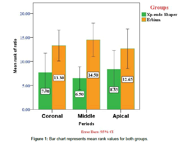
In Table 2, descriptive statistics between two groups within each site (apical, middle, and coronal) is shown. The mean rank values for both groups were showed in Figure 1. The data were collected and statistically analyzed using the Statistical Package for the Social Sciences (SPSS, version 21). Shapiro-wilk test: test the normality distribution of quantitative variables.
| Site | Groups | Min. | Max. | Mean | ±SD | Median | MR | Mann-Whitney U test | |
|---|---|---|---|---|---|---|---|---|---|
| Z | Sig. | ||||||||
| Apical | XP-endo Finisher | .000 | 81.460 | 32.008 | 31.797 | 24.450 | 8.35 | 1. 632 | 0.105 NS |
| Er:Cr:YSGG Laser | .000 | 100.000 | 59.118 | 34.324 | 66.955 | 12.65 | |||
| Middle | XP-endo Finisher | 10.420 | 77.950 | 54.560 | 28.446 | 70.215 | 6.50 | 3.089 | 0.002 HS |
| Er:Cr:YSGG Laser | 10.760 | 100.000 | 87.125 | 28.090 | 100.000 | 14.50 | |||
| Coronal | XP-endo Finisher | 20.220 | 100.000 | 68.202 | 26.672 | 75.140 | 7.70 | 2.187 | 0.035 S. |
| Er:Cr:YSGG Laser | 71.870 | 100.000 | 92.016 | 11.413 | 100.000 | 13.30 | |||
Table 2: Descriptive and statistical test of permeability among groups within sites.
Mann-Whitney U test the nonparametric test was done to determine whether there is a significant difference between groups regardless the level and between groups at different levels.
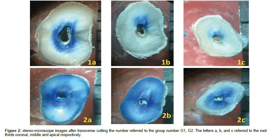
The results showed that there were a highly significant difference between Erbium laser group and XP-endo finisher group. For permeability test, stereomicroscopic images were taken after transversal cuts into three parts corresponding to root thirds as see in Figure 2. Scanning electron microscope micrographs were taken for each third (apical, middle and coronal) of specimens at 500 & 1000 x for both groups. In the first group were using the XP-endo finisher file, it can be noticed that a slightly removal of smear layer. Also, there are some opened dentinal tubules especially in middle third, however, it was seen a semilunar shape of most of the dentinal tubules, which mean that they are partially occluded especially in apical and coronal thirds (Figure 3a and 3b).
For Erbium laser group the smear layer was ablated and removed in most areas of the root specifically in middle and apical thirds. The apical third of the root canal dentin suffered from ablation more than other areas particularly the inter-tubular dentin rather than peritubular dentin. In coronal third still there are some of the dentinal tubules partially occluded (Figure 4a and 4b).
Discussion
After mechanical preparation of root canal, an amorphous, irregular layer known as the smear layer is formed on root canal walls. Possible harmful effects may occur if the smear layer is not removed during root canal treatment. The smear layer may act as a harbour for microorganisms after the instrumentation of an infected root canal space, so bacteria and bacterial by products can survive and infect the canal again.
The smear layer has also been shown to impede the penetration of intracranial medicaments and sealers into dentinal tubules and has the potential of compromising the seal of the root filling [13-15]. Syringe irrigation is a standard way for root canal irrigation, but this technique is not effective in the apical third of the root canal [16]. It seems to be difficult to completely remove smear layer, particularly in the apical third of the root because the smaller size of the apical third (compared with the other thirds) hinders the circulation and action of the irrigating solutions and vapor lock formation [17]. Therefore, this study aimed to see the effect of the Er:Cr:YSGG laser with Xp-endo finisher on root canal dentin permeability and smear layer removal.
The main effect of root canal irrigants is to clean the root canals during the enlarging and shaping process [16].
Previous studies have investigated the ability of different concentrations of EDTA in combination with NaOCl for smear layer removal currently, this is the most effective method and widely accepted [14,18-21].
The finding in this study regarding the results of XP-endo Finisher file in debris and smear layer removal can be attributed to its metallurgy. The manufactures of XP-endo Finisher files are dependent on the shape-memory principles of the NiTi alloy. The file is straight in its martensitic phase which is formed when it is cooled. When the file is subjected to the body temperature (the canal) it will convert its shape because of its shape-memory to the austenitic phase. It has been claimed by the manufacturer that the austenitic phase shape in the rotation mode permits the file to contact and clean areas that are otherwise difficult to reach with regular instruments. Even though neither the smear layer was not completely removed nor the dentinal tubules opened clearly compared with cavitation effect formed by laser activated irrigation [22].
For Erbium laser group vapour bubbles form when laser energy is absorbed by irrigant solution, which can cause a volume expansion that is 1600 times the original volume and then collapse and cause an acoustic streaming which in order, causes the cavitation effect [23].
Conclusion
The Er:Cr:YSGG laser ablative effect was clear especially in apical third. For that reason, we concluded that direct laser irradiation accompanied with cavitation effect done by laser activation of irrigants was the most effective protocol in removing smear layer and increase dentin permeability.
References
- Ferreira RB, Alfredo E, Arruda MP, Sousa YT, Sousa-Neto MD (2004) Histological Analysis Of The Cleaning Capacity Of Nickel-Titanium Rotary Instrumentation With Ultrasonic Irrigation In Root Canals. Australian Endodontic Journal 30: 56-58.
- McComb D, Smith DC (1975) A preliminary scanning electron microscopic study of root canals after endodontic procedures. Journal of endodontics 1: 238-242.
- Hulsmann M, Hahn W (2000) Complications during root canal irrigation–literature review and case reports. Int Endod J 33: 186-193.
- Huang TY, Gulabivala K, Ng YL (2008) A bio-molecular film ex-vivo model to evaluate the influence of canal dimensions and irrigation variables on the efficacy of irrigation Int Endod J 41: 60-71.
- McGill S, Gulabivala K, Mordan N, Ng YL (2008) The efficacy of dynamic irrigation using a commercially available system (RinsEndo®) determined by removal of a collagen ‘bio-molecular film’from an ex vivo model. Int Endod J 41: 602-608.
- Gu LS, Kim JR, Ling J, Choi KK, Pashley DH et al. (2009) Review of contemporary irrigant agitation techniques and devices. Journal of Endodontics. 35: 791-804.
- Matsumoto H, Yoshimine Y, Akamine A (2011) Visualization of irrigant flow and cavitation induced by Er: YAG laser within a root canal model. Journal of endodontics 37: 839-843.
- Levy G, Rizoiu I, Friedman S (1996) Lam H. Pressure waves in root canals induced by Nd: YAG laser. Journal of endodontics 22: 81-84.
- Cheng X, Guan S, Lu H, Zhao C, Chen X et al (2012) Evaluation of the bactericidal effect of Nd: YAG, Er: YAG, Er, Cr: YSGG laser radiation, and antimicrobial photodynamic therapy (aPDT) in experimentally infected root canals. Lasers in surgery and medicine 44: 824-831.
- Cheng X, Chen B, Qiu J, He W, Lv H (2016) Bactericidal effect of Er: YAG laser combined with sodium hypochlorite irrigation against Enterococcus faecalis deep inside dentinal tubules in experimentally infected root canals. Journal of medical microbiology 65: 176-187.
- Al-Karadaghi TS, Franzen R, Jawad HA, Gutknecht N (2015) Investigations of radicular dentin permeability and ultrastructural changes after irradiation with Er, Cr: YSGG laser and dual wavelength (2780 and 940 nm) laser. Lasers in medical science 30: 2115-221.
- Marchesan MA, Brugnera-Junior A, Souza-Gabriel AE, Correa-Silva SR, Sousa-Neto MD (2008) Ultrastructural analysis of root canal dentine irradiated with 980-nm diode laser energy at different parameters. Photomedicine and laser surgery 26: 235-240.
- Liang YH, Jiang LM, Jiang L, Chen XB, Liu YY et al. (2013) Radiographic healing after a root canal treatment performed in single-rooted teeth with and without ultrasonic activation of the irrigant: a randomized controlled trial. Journal of endodontics 39: 1218-1225.
- Garip Y, Sazak H, Gunday M, Hatipoglu S (2010) Evaluation of smear layer removal after use of a canal brush: an SEM study. Oral Surgery, Oral Medicine, Oral Pathology, Oral Radiology, and Endodontology 110: e62-66.
- Wang Z, Shen Y, Haapasalo M (2013) Effect of smear layer against disinfection protocols on Enterococcus faecalis–infected dentin. Journal of endodontics 39: 1395-1400.
- Peeters HH, Suardita K (2011) Efficacy of smear layer removal at the root tip by using ethylenediaminetetraacetic acid and erbium, chromium: yttrium, scandium, gallium garnet laser. Journal of endodontics. 37: 1585-1589.
- Arslan H, Ayrancı LB, Karatas E, TopçuoÄŸlu HS, Yavuz MS (2013) Effect of agitation of EDTA with 808-nanometer diode laser on removal of smear layer. Journal of endodontics 39: 1589-1592.
- Basrani B, Haapasalo M (2012) Update on endodontic irrigating solutions. Endodontic topics 27: 74-102.
- Prado M, Gusman H, Gomes BP, Simao RA (2011) Scanning electron microscopic investigation of the effectiveness of phosphoric acid in smear layer removal when compared with EDTA and citric acid. Journal of endodontics 37: 255-258.
- Carvalho AS, Camargo CH, Valera MC, Camargo SE, Mancini MN (2008) Smear layer removal by auxiliary chemical substances in biomechanical preparation: a scanning electron microscope study. Journal of endodontics 34: 1396-1400.
- Rodig T, Dollmann S, Konietschke F, Drebenstedt S, Hülsmann M (2010) Effectiveness of different irrigant agitation techniques on debris and smear layer removal in curved root canals: a scanning electron microscopy study. Journal of endodontics 36: 1983-1987.
- FKG Dentaire SA (2016) The XP Endo Finisher File Brochure. La Chaux-de-Fonds. Switzerland.
- Blanken J, De Moor RJ, Meire M, Verdaasdonk R (2009) Laser induced explosive vapor and cavitation resulting in effective irrigation of the root canal. Part 1: a visualization study. Lasers in surgery and medicine 41: 514-519.
 Spanish
  Chinese
  Russian
  German
  French
  Japanese
  Portuguese
  Hindi
Hindi 


