Research Article, J Food Nutr Disor Japan Vol: 9 Issue: 1
Cancer Therapeutic and Preventive Effects of Dried Seaweed (Porphyra yezoensis) Extract by Gut Immunity Activation
Hideaki Ichihara1, Masaki Okumura1,2, Takashi Doi2, Tatsuro Inano2, Koichi Goto1 and Yoko Matsumoto1*
1Division of Applied Life Science, Graduate School of Engineering, Sojo, University, Japan
*Corresponding Author : Yoko Matsumoto
Division of Applied Life Science, Graduate School of Engineering, Sojo University, 4-22-1 Ikeda, Nishi-Ku, Kumamoto 860-0082, Japan
Tel: +81-96-326-3965
E-mail: matumoto@life.sojou.ac.jp
Received: January 20, 2020 Accepted: January 28, 2020 Published: February 6, 2020
Citation: Ichihara H, Okumura M, Doi T, Inano T, Goto K, et al. (2020) Cancer Therapeutic and Preventive Effects of Dried Seaweed (Porphyra yezoensis) Extract by Gut Immunity Activation. J Food Nutr Disor 9:1.
Abstract
The application of active ingredients in biomass in pharmaceutical products is eagerly anticipated, such as the use of dried seaweed (nori, Porphyra yezoensis) currently discarded as biomass. In this study, we used mice subcutaneously transplanted with malignant melanoma and lymphoma cells as models to examine the cancer-preventive and therapeutic effects of dried seaweed extract mediated by the immuno-stimulatory effects of components of the extract. The 15-day oral administration of seaweed extract resulted in lower tumor (melanoma) weight in the model mice. Time-dependent increases in IgA and IgG levels were observed in the serum of melanoma model mice following oral administration of seaweed extract. We observed an increase in IgA levels in a solution of the supernatant from homogenized ileal tissue from the melanoma model mice following the administration of seaweed extract. Furthermore, immunostaining revealed multiple IgA-positive cells in the ileal tissue sections of the melanoma model mice that were orally administered seaweed extract. In addition, when the model mice were orally administered seaweed extract for 7 days, the size of the subcutaneous lymphoma tumors tended to decrease, suggesting the therapeutic effect of the extract. When the mice were administered seaweed extract as a pre-treatment for 7 days before lymphoma tumor transplantation, significant shrinkage of the tumor was observed in the seaweed extract-treated mice compared with the control mice, which suggested the preventive effect of seaweed extract.
Keywords: Porphyra yezoensis; Immunostimulation; Cancer therapeutic effect; Cancer preventive effects; Porphyrin
Introduction
Cancer is the most common pathological cause of death in the world. The number of deaths due to cancer is increasing each year, indicating the need to develop new treatments with a high success rate and few side effects. Cancer treatments can be classified into three major types: surgery, chemotherapy, and radiation therapy. However, despite the beneficial effects of these treatments, there are also negative effects involved. In recent years, immunotherapy has emerged as the fourth type of cancer treatment [1].
Recently, seaweed (nori, Porphyra yezoensis ) has attracted interest as food that improves the body’s immunity. Nori is a common food for Japanese people, who have a long history of eating algae. Seaweed contains approximately 30% porphyrin, a polysaccharide. Porphyrin is reported to have immunostimulatory effects [2,3]. However, despite the elucidation of this function of seaweed, the majority of seaweed is deemed to be of no value, and classed as scrap seaweed, owing to the discoloration that occurs during the cultivation process. Therefore, the application of seaweed to products with added value, such as biomass, is an area for exploration.
We have been researching the application of common materials for medical engineering. The short-term oral administration of seaweed extracts to mice bearing abdominal melanoma has been shown to increase IFN-γ levels [4].
In this study, we examined the potential of seaweed as a treatment for cancer through its in vivo immunostimulatory effects on mice bearing subcutaneously transplanted melanoma and lymphoma cells.
Material and Methods
Samples and component extraction
For seaweed (nori), we used a seaweed extract in which the main ingredient of Porphyra yezoensis harvested in the Ariake Sea (Seaweed porphyrin solution, purity: 80% +, Daiichi Seimo Co., Ltd., Fukuoka, Japan), which was spray-dried at Ohmoriya Co. Ltd. Osaka, Japan to obtain a powdered sample. The seaweed extract powder was dissolved in ultrapure (0.20-μm filtered) water. The mixture was sonicated (40°C, 1 mL/mL) until completely mixed, and then autoclaved.
Cells and animals
Mouse melanoma cells (B16-F0) were purchased from the DS pharma-bio medical Co., Ltd. Osaka, Japan and cultured in DMEM medium (GIBCO, Thermo Fisher Scientific, MA, USA) containing FBS. Mouse lymphoma cells (YAC-1) were purchased from the Institute of Physical and Chemical Research (RIKEN) and cultured in RPM1640 medium (GIBCO) containing 10% fetal bovine serum (FBS, Hyclone Laboratories, Inc., UT, USA). Five-week-old, female A/ JJmsSlc, and C57BL/6NJcl mice were purchased from Japan SCL (Shizuoka, Japan), Inc. and CLEA Japan, Inc. (Tokyo, Japan), respectively.
The therapeutic effect of seaweed extract following repeated oral administration in mice bearing subcutaneously transplanted melanoma
The mice were handled in accordance with the guidelines for animal experiments stipulated by Japanese law. The animal experiments were conducted following approval from the Animal Experiments Ethics Committee of the Sojo University. After 2 days of acclimation, the bodyweight of the C57BL/6NJcl mice was measured and the mice were allocated to the sample treatment groups or the control group by stratified randomization. The mice were housed in an environment with a room temperature of 24°C ± 2°C and humidity of 55% ± 10% and given free access to water and feed. B16-F0 melanoma cells (5.0 × 10⁶ cells/mouse) suspended in PBS was transplanted into the mice, which were then allocated to one of five groups: the control group (water) or one of the seaweed extract treatment groups (10 mg/kg, 25 mg/kg, 50 mg/kg, or 75 mg/kg). The mice were orally administered the samples for 15 days continuously starting from the date of transplantation. On day 16, the mice were dissected under anesthesia and the tumors were removed. We evaluated the anticancer effect of seaweed extract through observations of the general condition, the measurement of body weight, and the measurement of tumor weight.
Evaluation of the therapeutic effect over time of seaweed extract in mice bearing subcutaneously transplanted melanoma through immunostimulation
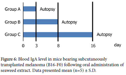
The B16-F0 melanoma cell solution (1.0 × 10⁶ cells/mouse) was subcutaneously transplanted using Matrigel. After transplantation, the mice were allocated into two groups (control (water) or 25 mg/kg bodyweight seaweed extract treatment) and orally administered samples from the date of transplantation. To measure the changes over time between Day 3 and Day 16, individual mice were selected at regular intervals and dissected. In group A, the mice were treated until Day 2 and dissected on Day 3 (five animals per group). In group B, the mice were treated until Day 7 and dissected on Day 8 (five animals per group). In group C, the mice were treated until Day 15 and dissected on Day 16 (five animals per group). During the period of oral administration, we measured the bodyweight of the mice and the tumor volume. On Days 3, 8, and 16, we collected serum samples from the dissected mice. The therapeutic effect of seaweed extract was evaluated with respect to the change in tumor weight (Figure 1).
Measurement of IgA, IgG, IFN-γ, and IL-12 levels using ELISA in mice bearing subcutaneously transplanted melanoma treated with seaweed extract
Blood samples were collected during the dissection procedure described in Section 1-4, in order to obtain a serum sample. The IgA, IgG, IFN-γ, and IL-12 levels in the serum samples were measured by using ELISA kits in accordance with the manufacturer’s protocols. ELISA measurements of IgA and IgG levels were performed by using a kit from Bethyl Laboratories Inc. (TX, USA), the IFN-γ and IL-12 levels were measured by using a kit from R&D Systems (MN, USA).
A sample of ileal tissue was taken, homogenized in PBS (200 μL), and the supernatant was collected. The IgA levels in the homogenized supernatant and serum samples were measured by using the Mouse IgA ELISA Kit (Bethyl Laboratories) in accordance with the manufacturer’s protocol.
IgA immunostaining of ileal tissue sections
On Day 16, as described in Section 2-4, the mice were dissected under anesthesia, and a sample of ileal tissue was taken. The tissue sample was fixed in formalin solution, immunostained with IgA antibodies, and then stained with 3,3'-Diaminobenzidine Tetrahydrochloride (DAB). The stained tissue sections were observed using an optical microscope (TS-100, Nikon Corporation, Tokyo, Japan).
Study of the therapeutic effect of seaweed extract in mice subcutaneously transplanted with YAC-1 cells
The mice were divided into two treatment groups (control (water) and 25 mg/mL seaweed extract) and received subcutaneous transplantation of YAC-1 cell suspension (5.0 × 10⁶ cells/body) suspended in PBS (-). Subsequently, the mice were treated for 7 days from the date of transplantation (Day 1), with the bodyweight of the mice measured throughout the treatment period. On day 8, the mice were dissected and the tumor weight was measured; this was used to evaluate the therapeutic effect.
Study of the cancer-preventive effect of seaweed extract in mice subcutaneously transplanted with YAC-1 cells
The mice were divided into two treatment groups (control or 25 mg/mL seaweed extract) and were orally administered the treatment for 7 days. The bodyweight of the mice was measured during this period. After treatment for 7 days, a YAC-1 cell suspension (5.0 × 10⁶ cells/mouse) suspended in PBS (-) was subcutaneously transplanted into the mice (Day 1). The bodyweight of the mice was measured for 7 days after transplantation. On Day 8 after transplantation, the mice were dissected and the tumor weight was measured; this was used to evaluate the preventive effect.
Statistical analysis
Data were statistically analysed by a Student’s t-test. All data are presented as the mean ± SD. p values less than 0.05 were considered to be statistically significant.
Results and Discussion
Therapeutic effect of seaweed extract following its repeated oral administration in mice bearing subcutaneously transplanted melanoma
Fucoidan, a polysaccharide combined with sulfuric acid and uronic acid, is a viscous substance that can be extracted with water or dilute hydrochloric acid. In recent years, interest in its ability to induce apoptosis in cancer cells has increased; in addition, it is known to increase the production of interleukin and interferon, which are compounds with therapeutic action against allergy and cancer [5].
Alginic acid and fucoidan are typical polysaccharides contained in Kombu (edible kelp) that act to control cancer; fucoidan comprises 40% to 60% of the content of Kombu [6].
With respect to the immunostimulatory effect of seaweed, it is known that the administration of fucoidan to mice activates antigenpresenting cells such as macrophages, which leads to enhanced production of IL-12, IFN-ɣ production by T-cells, and the activation of NK cells. It has also been reported that the porphyrin and porphyrinderived oligosaccharides contained in seaweed activate the mouse immune system [4,7].
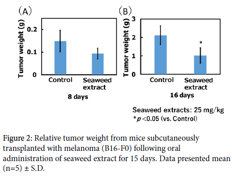
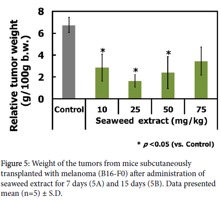
In this study, we examined the therapeutic effect of orally administered seaweed extract in mice bearing subcutaneously transplanted tumors. First, we examined the therapeutic effect of the substance following repeated administration of the 10 mg/kg, 25 mg/kg, 50 mg/kg, and 75 mg/kg seaweed extract for 15 days to the C57BL/6 mice subcutaneously transplanted with B16-F0 melanoma cells. There were no abnormal changes in the bodyweight of mice during the experimental period (data not shown). There were no abnormalities in the bodyweight of mice during the treatment period. The relative tumor weight is shown in Figure 2.
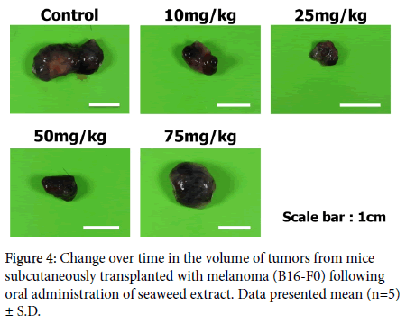
There was a significant decrease in tumor weight in all groups compared with the control group, which provided a clear indication of the therapeutic effect of seaweed extract. Photographs of the tumors removed from each treatment group are shown in Figure 3. The tumors in the sample groups had shrunk compared to those in the control group. We observed a therapeutic effect at a concentration below 75 mg/kg, which was close to the 50 mg/kg dose at which the therapeutic effect was observed in a previous therapeutic experiment.
In particular, a significant reduction in tumor weight was observed in the 25 mg/kg group. The measurement of IFN-γ levels by ELISA did not detect increases after any treatment. The above results confirmed the tumor-shrinking effect of seaweed extract has on B16-F0 melanoma tumors.
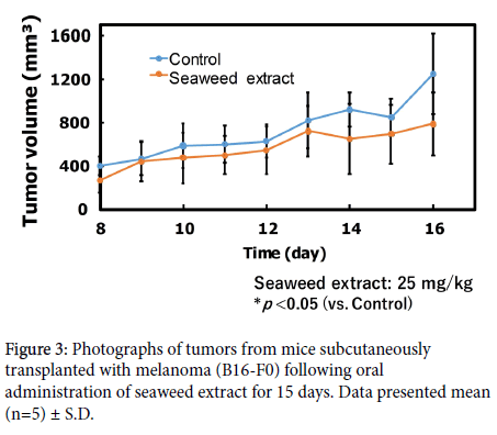
Furthermore, we examined the therapeutic effect of seaweed extract over time following repeated oral administration of 25 mg/kg seaweed extract for 2 (group A), 7 (group B), and 15 (group C) days to C57BL/6 mice bearing subcutaneously transplanted B16-F0 melanoma cells. There were no abnormal changes in the bodyweight of mice during the treatment period (data not shown). There were no major changes in the bodyweight of mice in either the untreated group or the seaweed extract-treated group, which indicated the absence of toxicity. The tumor size measurements during the treatment period are shown in Figure 4.
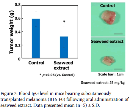
In the seaweed extract-treated group, the growth of the tumor volume was smaller than that of the growth in the untreated group. The tumor weight of tumor-bearing model mice that were dissected on Day 8 is shown in Figure 5. On Day 8, we confirmed the shrinkage of tumors in the seaweed extract-treated groups relative to the untreated group. The tumor weight of the tumor-bearing model mice dissected on Day 16 is shown in Figure 5. Significant shrinkage (p<0.05) was found in the tumors of mice in the seaweed extract-treated group compared with the untreated group, thereby elucidating the therapeutic effect of seaweed extract.
In summary, the mice administered seaweed extract showed shrinkage in tumor weight after 8 days, and significant shrinkage after 16 days.
The immunostimulatory effect of seaweed extract following oral administration to mice subcutaneously transplanted with melanoma
Immunostimulation refers to an action that activates the body’s immunity, enhancing the defensive power of the body, enabling the body to protect itself from foreign substances. The human immune mechanism can be broadly divided into “ innate immunity ” and “acquired immunity”. Innate immunity is the first type of immunity to respond to external threats. The human body has immune cells, such as NK cells, which are able to attack viruses and bacteria that come from outside the body. In addition to NK cells, innate immunity involves the action of substances called immunoglobulin A (IgA). IgA normally functions as neutralizing antibodies in the body; their role in innate immunity is to coating antigens, preventing any toxins released by the antigens from spreading around the body. Acquired immunity works through the specific identification and memorization of an infective pathogen, which allows its effective removal by the immune system upon future exposure to the pathogen) [8].
IgA is produced by plasma cells present in the intestines and is abundant in digestive secretions, saliva, the respiratory tract mucosa, and breast milk. Secreted IgA works with lysozymes to break down the sugar chains in the bacterial cell walls, thereby killing the bacteria. As IgA reacts to a variety of pathogens, rather than reacting to specific viruses and bacteria, it is known to be effective against a wide range of pathogens [9-11].
In addition, an indispensable part of acquired immunity is immunoglobulin G (IgG). IgG is abundant in the blood, lymph fluids, and bone marrow, and is the most common type of immunoglobulin found in the blood. IgG reacts with macrophages, neutrophils, and NK cells to activate the complement system. Owing to its long lifetime in the blood, IgG is a key factor in immunity. It is also important in immunology and clinical diagnosis [12].
In this study, we examined the immunostimulatory effect of seaweed extract following oral administration to tumor-bearing mice.
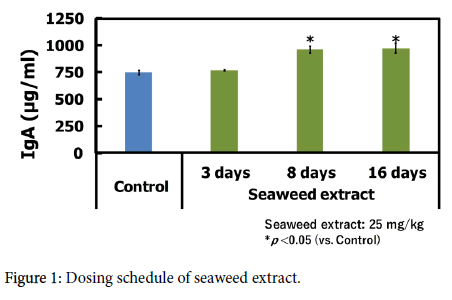
First, we measured the IgA, IgG, IFN-ɣ and IL-12 levels in the mouse serum by using ELISA. The IgA levels in tumor-bearing mice that were administered seaweed extract are shown in Figure 6. Compared with the untreated mice, the seaweed extract-treated mice showed increased IgA production, with a significant increase observed on Day 8 and Day 16. IgG production tended to increase in the seaweed extract-treated groups throughout the treatment period, but with no significant difference to the untreated group (Figure 7). In contrast, the IFN-ɣ and IL-12 levels were similar to those in the untreated group (data not shown). These results confirmed the significant increase in IgA and a slight increase in IgG in the treatment group, which suggested that immunostimulation by seaweed extract may be an effective cancer treatment.
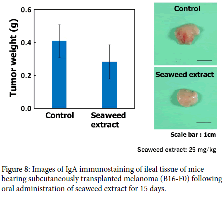
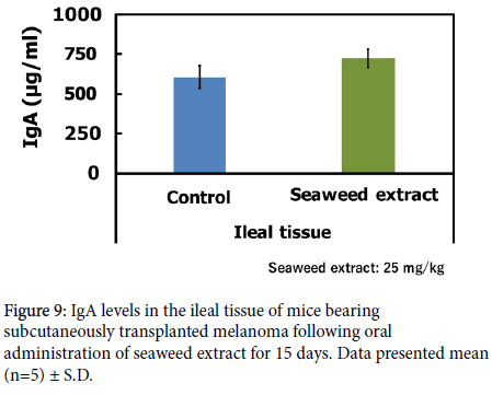
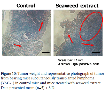
Next, we performed IgA immunostaining of ileal tissue sections from the tumor-bearing mice. After 15 days of administering the seaweed extract, the animals were dissected on Day 16 under anesthesia, in order to prepare tissue sections. The tissue sections were then immunostained using IgA antibodies and observed under the optical microscope. The results are shown in Figure 8. In all samples, we observed brown-stained IgA-expressing cells. The seaweed extracttreated group showed more IgA-expressing cells near Peyer’s patches than the untreated group. Furthermore, we measured IgA levels in the homogenized supernatant of the ileal tissue from tumor-bearing mice that received repeated administration of seaweed extract; the results are shown in Figure 9. The IgA levels in the ileum and serum were increased in tumor-bearing mice that received repeated oral administration of seaweed extract. These results suggested the involvement of immunostimulation in the tumor-shrinking effect of seaweed on B16-F0 melanoma tumors, which was potentiated by the function of IgA in intestinal immunity.
Immunostimulatory effect of seaweed extract following oral administration to lymphoma-bearing mice
From the date of transplanting lymphoma (YAC-1) cells, the mice were orally administered seaweed extract (Dose: 25 mg/mL) for 7 days. On Day 8 after transplantation, the mice were dissected to have the tumor weight weighed and to evaluate the therapeutic effect of seaweed extract. We evaluated the therapeutic effect of seaweed extract on lymphoma-bearing mice and found no abnormal changes in body weight during the treatment period in the seaweed extract-treated mice.
There was no significant difference in the tumor weight between the seaweed extract-treated group and the control group (Figure 10). However, there the tumor weight tended to be lower in the seaweed extract-treated group, which suggested the therapeutic effect of seaweed extract. The photographs also suggested a decrease in tumor size following the administration of seaweed extract.
After orally administering seaweed extract (Dose: 25 mg/mL) for 7 days, the mice were transplanted lymphoma (YAC-1) cells. On Day 8 after transplantation, the tumor weight was measured in order to evaluate the therapeutic effect of seaweed extract. We also evaluated the preventive effect of seaweed extract on mice subcutaneously transplanted with lymphoma cells.
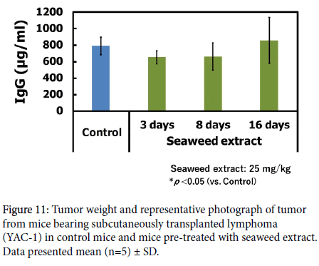
On Day 8 after transplantation, compared with the control group, a significant decrease was observed in tumor weight in the seaweedtreated group, demonstrating the preventive effect of pre-treatment with seaweed extract (Figure 11). Photographs also indicated smaller tumors in the seaweed extract-treated group than in the control group.
As shown by these results, the repeated oral administration of seaweed extract activated intestinal immunity in Peyer’s patches and other areas, mediating preventative and therapeutic effects by strengthening the immune response of mice to the subcutaneously transplanted cancer.
Conclusion
This study examined the anticancer effect of dried seaweed extract resulting from its immunostimulatory properties, which may support an effective use for dried seaweed that will otherwise be discarded as bioweight. The following findings were determined in our study:
The 15-day repeated oral administration of seaweed extract (25 mg/kg) in mice subcutaneously transplanted with melanoma (B16-F0) resulted in a significant decrease in tumor size.
The 15-day repeated oral administration of seaweed extract (25 mg/kg) in mice subcutaneously transplanted with melanoma (B16-F0) resulted in higher IgA and IgG levels in mouse blood over time.
The 15-day repeated oral administration of seaweed extract (25 mg/kg) in mice subcutaneously transplanted with melanoma (B16-F0) resulted in higher in IgA levels in the ileal tissue of mice, as shown by ELISA and immunostaining.
The 7-day repeated oral administration of seaweed extract (25 mg/kg) to mice subcutaneously transplanted with lymphoma (YAC-1) cells resulted in a reduction in tumor size, confirming the therapeutic effect of seaweed extract.
When lymphoma (YAC-1) cells were subcutaneously transplanted to mice that received a 7-day pre-treatment of the oral administration of 25 mg/kg seaweed extract, the tumor size decreased, showing the preventative effect of seaweed extract.
We have elucidated the profound anticancer effect of seaweed extract in vivo through immunostimulation, with the suggested involvement of intestinal immunity. In the future, seaweed extract may be developed as a natural health food substance.
Disclosure Statement
No potential conflict of interest was reported by the authors.
References
- National Cancer Center (2018) Cancer Information Services (Immunotherapy).
- Yoshizawa Y, Enomoto A, Todoh H, Ametani A, Kaminogawa S (1993) Activation of murine macrophages by polysaccharide fractions from marine algae (Porphyra yezoensis). Biosci Biotech Biochem 57: 1862-1866.
- Osumi Y, Kawai M, Amano H, Noda H (1998) Antitumor activity of oligosaccharides derived from porphyra yezoensis porphyran. Nippon Suisan Gakkaishi 64: 847-853.
- Kitajima H, Komizu T, Ichihara H, Goto K, Doi T, et al. (2013) Immunostimulatory effects of extract from Nori (Porphyra yezoensis) in vitro and in vivo. Kagaku Kougaku Ronbunshu 39: 359-362.
- Yamada N (2000) The chemistry of using seaweed. Naruyamado Shoten 9: 88-90.
- Notoya M (2000) Biology of nori. Naruyamado Shoten, p: 148.
- Kenneth M, Paul T, Mark W (2010) Immunobiology. Nankodo Shoten, pp: 469-837.
- Japan Blood Products Association (2018) Immunoglobulin.
- Otsuka Pharmaceutical (2018) Pharmaceutical: IgA antibody. www.otsuka.co.jp/b240/m/mechanism.html.
- You-Gui L, Dong-Feng J, Shi Z, Jian-Xun Z, Shi C, et al. (2011) Anti-tumor effects of proteoglycan from Phellinus linteus by immunomodulating and inhibiting Reg IV/EGFR/Akt signaling pathway in colorectal carcinoma. Int J Biol Macromol 48: 511-517.
- Easy tips on increasing your immunity (2016) Shufunotomo Inc, pp: 110-112.
- Miyazaki Y, Kirino T, Yamaguchi S (2014) Small-scale clinical study related to immune potentiation and safety of fucoidan-containing food items-on the improvement of oral mucosa immune mechanism. Food Style 18: 21-25.
 Spanish
  Chinese
  Russian
  German
  French
  Japanese
  Portuguese
  Hindi
Hindi 