Spanish
  Chinese
  Russian
  German
  French
  Japanese
  Portuguese
  Hindi
Hindi Research Article, J Food Nutr Disor Vol: 8 Issue: 3
Comparative Effects of Garlic, Yogurt, Beniseed Liquor and Fresh Orange Juice on Induced Type-1 Diabetes Mellitus in Rabbits using Streptozotocin
1Department of Microbiology, Federal University of Technology, Akure, P.M.B. 704, Akure, Ondo State, Nigeria
2Department of Biological Sciences, Elizade University, Ilara-Mokin, Ondo State, Nigeria
*Corresponding Author : Momoh AO
Department of Biological Sciences, Elizade University, Ilara-Mokin, Ondo State, Nigeria
Tel: +234803 6530 924
E-mail: abdul.momoh@elizadeuniversity.edu.ng
Received: September 11, 2019 Accepted: October 11, 2019 Published: October 21, 2019
Citation: Omoya FO, Momoh AO (2019) Comparative Effects of Garlic, Yogurt, Beniseed Liquor and Fresh Orange Juice on Induced Type-1 Diabetes Mellitus in Rabbits using Streptozotocin. J Food Nutr Disor 8:4.
Abstract
Streptozotocin (STZ) was used to induce type-1 diabetes mellitus in animal models (twenty-one rabbits) at 60 mg/kg birth weight with two weekly booster doses to cause chronic diabetes in New Zealand White rabbits. They were given single intravenous dose of STZ in 1mL citrate buffer having a pH 4.6 and the booster dose given after 7 days and 14 days respectively. The blood glucose level was monitored along with the clinical signs including changes in behavior and weight loss. Treatment commenced immediately after the third booster when the rise in blood sugar was observed (4 hours later). They were treated with glucovance (a drug), fresh garlic extract, yogurt, beniseed liquor and orange juice for a period of 12 weeks (3 months) with the effect of the treatments checked on the glucose level of their blood, full blood count analysis using Mindray BC3300 auto-hematology analyzer and histopathology analysis of their pancreas. The results showed that the glucose level of the rabbits was within 70.02 ± 1.0 mg/dl at p ≤ 0.05 before they were induced. The level rose to 187.33 ± 0.9 mg/dl after the 3rd booster dose of streptozotocin was given to the rabbits. Gross morphology of selected organs showed that the inducement caused discoloration of the kidneys, oedema of the pancreas and significant increase in weight of the heart at p ≤ 0.05. There were no significant differences at p ≤ 0.05 between Packed Cell Volume (PCV) of the control group and the group induced and treated with diabetes drug; while there were significant differences in the fibrinogen values for the experimental groups. The induced type 1 diabetes mellitus caused an increase in the basophils, monocytes, and neutrophils while it caused significant decrease in the percentage of lymphocytes. Histopathologically, the inducement caused poor formation of the islet of Langerhans cells and dot necrotized cells. There is profuse hemorrhage from highly vascularized pancreatic acini surrounded by parenchyma fat cells. The group induced and treated with garlic extract showed some good features that not only depict a good and fast recovery from diabetes but showed some other health benefits of garlic. They are presence of well-formed pancreatic acini and cell infiltrations with well-spaced interstitial cells of the pancreas that shows active cell division of the pancreatic ducts and acini. There is splay and intrafaradization of the cracked pancreatic ducts. The presence of artifacts is seen at the anterior portion of the plate with well-formed interlobular and intralobular ducts. The effect of garlic extract as a good antidiabetic agent has been well established in this research. The curative strength of other foods such as yogurt and fruit extract has been proven in the findings of this research. Therefore, garlic has the most outstanding positive effect on diabetes and is recommended for people who are diabetic to feed on it or use it as food supplement.
Keywords: Type-1 diabetes; Streptozotocin; Garlic extract; Yogurt; Orange juice; Beniseed liquor
Introduction
The domestic rabbit (Oryctolagus cuniculus) is a multi-purpose animal kept in captivity for a different purpose (meat source, breeding stock, laboratory animal, pet, wool source and ornamental [1]. The New Zealand white rabbits are unique with special features. They are small mammals with white fluffy, short tails and whiskers as well as distinctive long ears. There are other species of rabbits all over the world in which are found in many different environments but they all have many things in common. They are herbivores, social creatures and live in large groups called colonies. The busiest time of day for rabbits is at dusk and dawn.
Diabetes may best be described as a group of metabolic disorders in which there are high blood sugar levels over a prolonged period. Symptoms of this disease are majorly weight loss, thirst, frequent urination, and increased hunger [2]. If left untreated, diabetes can cause many complications. Diabetes mellitus is a chronic, lifelong condition that affects the body’s ability to use the energy found in food [3]. There are three major types of diabetes: type 1 diabetes, type 2 diabetes, and gestational diabetes.
All types of diabetes mellitus have something in common, the cells need insulin, a hormone, in the bloodstream in order to take in the glucose and use it for energy. With diabetes mellitus, either the body doesn’t make enough insulin; it can’t use the insulin it does produce, or a combination of both [2]. This causes glucose to build up in the blood. High levels of blood glucose can damage the tiny blood vessels in the kidneys, heart, eyes, or nervous system; eventually causing heart disease, stroke, kidney disease, blindness, and nerve damage.
Insulin sensitizers are drugs that make the body much more sensitive to the action of insulin produced by the body or given as injection thereby helping to control the blood sugar level. Glucovance contains a combination of glyburide and metformin. Glyburide and metformin are both oral diabetes medicines that help control blood sugar levels [2].
Studies have shown that garlic and garlic extract offer lots of benefits to diabetes patients and has served both therapeutic and prophylactic measure in the control as well as prevention of diabetes [4]. Equally, sugar levels in fruit juice have been argued to cause a significant spike in blood sugar levels, increasing the risk of hyperglycemia (too high blood sugar levels). The glycemic index, which is used to reflect the impact on blood sugar levels of individual foods, places orange juice between 66 and 76 on a scale of 100.
However, eating the whole fruit instead of fruit juice has been said to give better nutrition and blood sugar management [5]. A whole medium orange contains 3 to 4 grams of fiber, compared with barely 1 gram in 8 ounces of juice. Fiber slows digestion time (so a whole orange won’t raise your blood sugar as quickly as juice), increases fullness, contributes to normal bowel function, and is associated with a decreased risk of many chronic diseases [6].
Yogurt, on the other hand, is a drink most people take either for pleasure, occasions or to quench thirst. Low-fat yogurt naturally contains both high-quality carbohydrates and protein, making it an excellent food for slowing or preventing an unhealthy rise in blood sugar. Studies also show that a diet high in calcium from yogurt and other calcium-rich foods is associated with a reduced risk of type 2 diabetes.
Previous research on beniseed, especially the fermented liquor has been proved to have immunostimulatory potential as well as the therapeutic effect on infectious diseases [4]. Toxicologically, the in vivo analyses on beniseeds revealed no deleterious effect on the organs of the rats. It has equally been recommended that for maximum result; fermenting beniseeds kept at 4°C ± 2°C is most effective [7]. Since yogurt has a much lower impact on glucose levels than people think, and raw or cooked garlic or aged garlic has been used to regulate blood glucose.
This research became necessary due to the current trend of diabetes which is on the rise as well as the fact that most of the drugs used for the treatment of the disease have severe side effects. Equally, most of the people that are diabetic in Nigeria and in Africa in general still prefer the use of herbs and foods to treat the disease than the use of modern drugs. Also, people have argued the effectiveness of the use of orange juice, unsweetened yogurts and other local preparations such as ogi liquor, beniseed liquor and even garlic in combating the terminal disease.
Therefore, this research aims to compare the effects of garlic juice, orange juice, yogurt and fermented beniseed liquor on induced type 1diabetes in rabbits.
Materials and Methods
Materials
The materials used in this research are: rabbits, wooden hutches, streptozotocin, needle and syringe, sterile distilled water, glucometer, top-load weight scale, auto-hematology analyzer, garlic juice, orange juice, yogurt, fermented beniseed liquor, a diabetes drug, etc.
Collection of rabbits
New Zealand White was purchased at 6-9 months old from a breeder farm and kept in separate hutches made of wooden materials. The hutches were properly disinfected before the arrival of the rabbits. They were allowed three weeks to adapt to the new environment. They were dewormed and given necessary medications during the experimental period. Fresh and green leaves of Tridax procumbens, Talinium triangulae and Aspergillia africana were given, while pelletized feeds was supplemented at regular intervals. Freshwater was given ad libitum.
The Department of Animal Production and Health as well as the unit of Animal Husbandry of the Federal University of Technology, Akure examined the health status of the rabbits to ensure they were apparently healthy. The experimental protocols were approved by the Animal Ethics Committee, of the Ondo State Veterinary Sciences and Animal Husbandry. The protocols were ensured to conform to the guidelines for the Care and Use of Laboratory Animals.
Study design
A total of twenty-one (21) young adult male rabbits were grouped into seven (7) groups of three (3) rabbits each. The weights of the rabbits were taken using top loading scale and special attention was taken to group rabbits of the same or near the same weight to be in the same group. They were allowed to acclimatize to the new environment for four weeks before inducing them.
Inducing type I diabetes
Streptozotocin (Sigma-Aldrich) was used at the dose rate of 60 mg/kg body weight, with two weeks consecutive booster doses based on available literature to attain hyperglycemia. The solution of the calculated dose for each animal was prepared in 1mL freshly prepared citrate buffer pH 4.6 (100 mM citric acid and 100 mM sodium citrate) immediately before use. Rabbits fasted for 18 hours followed by administration of slow intravenous injection of STZ through ear vein using insulin syringe.
Weight determination
The weight of the experimental rabbits was determined using Camry Premium Weighing Scale Model 40021 made in China.
Clinical signs monitored
Rabbits were monitored for clinical signs including changes in behavior, weight loss, appearance, activity, water/feed intake, urination/defecation or any other deviation. The blood glucose levels were recorded and the booster dose was given after 7 days and 14 days respectively. Treatment commenced immediately after the third booster when the rise in blood sugar was observed (4 hours later).
Treatments
The twenty-one (21) rabbits were grouped into 7groups comprising of 3 rabbits each per group. Group 1: Control; Group 2: Induced and treated with drug (Glucovance); Group 3: Induced and not treated; Group 4: Induced and treated with garlic juice; Group 5: Induced and treated with orange juice; Group 6: Induced and treated with yogurt; Group 7: Induced and treated with fermented beniseed liquor. With the exception of the group treated with the drug which was according to the dosage, all other groups were given two (2) ml of the substance twice daily for a period of 12 weeks (3 months).
Determination of blood glucose
Blood samples were collected from the central auricular artery using insulin syringe and three drops of blood immediately transferred on a clean and dry glass slide for blood glucose estimation using glucometer (Accu-Chek, Roche Diagnostics India Pvt. Ltd., Mumbai). The mean of the three readings was recorded as blood glucose level for the sample.
Haematological tests
Haematological tests such as Packed Cell Volume (PCV), Haemoglobin Concentration (HB), Red Blood Cell Count (RBC), Erythrocyte Sedimentation Rate (ESR), White Blood Cell Count (WBC), Mean cell volume (MCV), Mean cell haemoglobin (MCH), Mean cell haemoglobin concentration (MCHC) and White Blood Cell differential count (Lymphocytes, Neutrophils, Monocytes, Eosinophils, and Basophils) were done according to Cheesbrough [8] using Mindray BC3300 (2016 Model) auto-haematology analyzer.
Histopathological tests
Histopathological test on the pancreas was performed according to the methods of Baker et al. [9] and Cheesbrough [8] in the following process:
Fixation: Fixation of the tissue was done to prevent further enzymatic activity that usually leads to post-mortem autolysis. It also hardens tissue as well as kills microbes and keeps the tissue in its original form. The organs of the animals were collected and fixed in 10% buffered formalin.
Trimming: After fixation, the organs were trimmed to about 1-2 cm before dehydration.
Dehydration: Dehydration was done by passing the tissue through different concentrations of ethanol. It was done by the use of automatic tissue processor. They were dehydrated in graded percentages (50%, 70%, 80% and 100%) of ethanol for 1 and a half hour each at 30°C ± 2°C.
Clearing: Clearing of dehydrated tissue was done using 100% xylene. The tissues were left for 2 hours to remove any remnant alcohol completely.
Embedding: This was the placing of the cleared tissue in melted paraffin and allowed to harden. The tissues were left in the molten paraffin wax for 2 hours to embed properly.
Sectioning: This was done using a microtome. The tissues were sectioned to about 3-10 μ and floated in water bath at 37°C.
Hydration: This is the process of passing the tissue through water by passing it through different concentrations of alcohol. It was passed through xylene, 100%, 90%, 80%, 70% and 50% of ethanol for 11/2 hours at each concentration.
Staining: This was done using hematoxylin and eosin (H and E) stains. Haematoxylin stains the nucleus blue while eosin stains the cytoplasm acidophilic.
Dehydration, fixing and microscopy of stained slides: Dehydration was done again by passing the tissue through different concentrations of ethanol. It was done by the use of automatic tissue processor. The tissues were dehydrated in different percentages (50%, 70%, 80%, and 100%) of ethanol for 1 and a half hour each and cleared with xylene. The method of Cheesbrough [8] was used to remove excess stain under tap water. After clearing in xylene, the fixed tissue on a glass slide was fixed with Canada balsam and covered with coverslips [10]. The preparations were left in the oven at 40°C and then placed under the photo-microscope for examination [11].
Statistical analysis
Data obtained were subjected to descriptive one-way analyses of variance using SPSS version 22 and Duncan New multiple range tests were used as follow up test.
Results
The effect of the induced type 1 diabetes on the glucose level as treatment progresses is shown in Table 1. Week 0 shows the sugar level of the various groups before they were induced with streptozotocin. There were no significant differences in the result obtained for the various groups at p ≤ 0.05 as they all have almost the same values of 70.02 ± 1.0 mg/dl except for groups 1 and 5 that had 71.13 ± 0.3 mg/dl. However, after the inducement with the booster dose of the streptozotocin in week one, the sugar level shoots up drastically with the results exceeding 187.33 ± 0.9 mg/dl and all the groups significantly different at p ≤ 0.05. The control group was however almost constant in the value of the blood sugar with little fluctuations around 71.13 ± 0.3 mg/dl for the twelve weeks of the research. The other experimental groups showed variableness in the sugar levels at a different stage or week of the experiment. The group induced and treated with garlic extract is most significant among the experimental groups. Before the inducement, the sugar level of the group was 70.10 ± 0.2 mg/dl. The level rose to 187.33 ± 0.9 mg/dl after the third booster dose of streptozotocin was given to the rabbits. However, by the twelfth week of treatment with the garlic extract, the sugar level dropped to 74.12 ± 4.0 mg/dl.
| 1 | 2 | 3 | 4 | 5 | 6 | 7 | |
|---|---|---|---|---|---|---|---|
| Week 0 | 71.13 ± 0.3 b | 70.13 ± 5.1 a | 70.02 ± 1.0a | 70.10 ± 0.2 a | 71.11 ± 2.7 b | 70.00 ± 0.0 a | 70.22 ± 0.8 a |
| Week 1 | 73.09 ± 0.7 a | 190.17 ± 0.9 e | 184.40 ± 2.0 b | 187.33 ± 0.9 d | 190.20 ± 0.8 e | 186.08 ± 1.6 c | 191.39 ± 4.3 f |
| Week 2 | 70.02 ± 2.4 a | 180.20 ± 0.3 b | 186.06 ± 0.8 d | 181.13 ± 5.3 c | 188.12 ± 2.6 e | 186.10 ± 0.4 d | 180.08 ± 1.0 b |
| Week 3 | 72.12 ± 0.8 a | 175.05 ± 3.5 b | 183.13 ± 0.9 d | 174.12 ± 2.2 b | 174.06 ± 0.8 b | 180.22 ± 0.8 c | 175.15 ± 3.5 b |
| Week 4 | 72.80 ± 2.6 a | 166.30 ± 1.3 c | 180.02 ± 1.4 f | 150.06 ± 0.4 b | 171.13 ± 3.1 e | 181.21 ± 1.1 f | 169.15 ± 1.5 d |
| Week 5 | 74.20 ± 0.6 a | 153.13 ± 0.7 c | 181.11 ± 1.1 g | 132.18 ± 1.6 b | 168.10 ± 2.4 e | 174.20 ± 2.4 f | 165.23 ± 1.3 d |
| Week 6 | 71.62 ± 1.4 a | 150.20 ± 0.5 c | 179.91 ± 0.7 g | 124.22 ± 4.2 b | 167.13 ± 1.7 e | 172.14 ± 2.2 f | 153.13 ± 1.7 d |
| Week 7 | 75.05 ± 3.5 a | 130.10 ± 0.2 c | 180.00 ± 0.0 g | 105.05 ± 0.5 b | 164.12 ± 0.4 e | 171.13 ± 5.3 f | 151.51 ± 3.9 d |
| Week 8 | 73.13 ± 1.7 a | 121.11 ± 1.3 c | 177.13 ± 0.3 g | 98.08 ± 0.8 b | 162.34 ± 0.8 e | 170.10 ± 2.0 f | 150.20 ± 1.2 d |
| Week 9 | 72.02 ± 2.2 a | 102.18 ± 6.2 c | 175.15 ± 0.5 g | 92.28 ± 0.4 b | 161.13 ± 0.7 e | 164.18 ± 0.6 f | 155.15 ± 5.1 d |
| Week 10 | 75.55 ± 3.5 a | 79.47 ± 0.7 b | 178.26 ± 1.2 f | 84.02 ± 0.4 c | 156.08 ± 3.6 e | 155.15 ± 1.9 d | 153.17 ± 2.3 d |
| Week 11 | 71.31 ± 1.3 a | 71.31 ± 0.5 a | 175.55 ± 2.5 e | 79.19 ± 3.1 b | 155.05 ± 1.5 c | 160.06 ± 0.2 d | 154.06 ± 0.8 c |
| Week 12 | 71.19 ± 0.5 b | 68.22 ± 4.2 a | 174.04 ± 2.6 f | 74.12 ± 4.0 c | 153.33 ± 3.9 d | 160.24 ± 0.6 e | 151.33 ± 3.3 d |
Keys: Group 1: Control; Group 2: Induced and treated with drug; Group 3: Induced and not treated; Group 4: Induced and treated with garlic juice; Group 5: Induced and treated with orange juice; Group 6: Induced and treated with yoghurt; Group 7: Induced and treated with fermented beniseed liquor
Table 1: The effect of the induced diabetes on the glucose level (mg/dl) as treatment continued.
Table 2 shows the effect of the induced type-1 diabetes on the gross morphology and weight of selected vital organs of the experimental rabbits. The gross morphology of the organs in groups 1 and 4 are all normal without any form of swelling or edema. Although, the organs in group 2 are normal; the kidneys are a bit whitish in color. The pancreas of groups 5, 6 and 7 are enlarged in size. The weight of the heart showed that while the control group was 2.1 ± 0.2 g, this value was significantly different from the values obtained for groups 4 and 5 (2.4 ± 0.6 g) both of which showed no significant differences at p ≤ 0.05. The results of the weight of the other organs (liver, kidneys and the pancreas) are captured in Table 2.
| Group | Description of gross morphology | Heart | Liver | Kidneys | Pancreas |
|---|---|---|---|---|---|
| 1 | All normal | 2.1 ± 0.2a | 1.3 ± 0..5 a | 0.4 ± 0.1 a | 0.1 ± 0.1 a |
| 2 | Kidneys are milky white, others are normal | 2.3 ± 0.7b | 1.7 ± 0.5 d | 0.7 ± 0.1 d | 0.13 ± 0.1 b |
| 3 | Kidneys are reddish brown, pancreas is enlarged and liver is light red | 2.5 ± 0.3 d | 2.2 ± 0.1 e | 0.9 ± 0.1 e | 0.32 ± 0.1 e |
| 4 | All normal | 2.4 ± 0.6 c | 1.4 ± 0.2 b | 0.5 ± 0.1 b | 0.12 ± 0.1 b |
| 5 | The heart, kidneys and pancreas are enlarged | 2.4 ± 0.4 c | 1.6 ± 0.2 c | 0.7 ± 0.1 d | 0.22 ± 0.1 d |
| 6 | The liver and pancreas are enlarged | 2.5 ± 0.5 d | 1.6 ± 0.4 c | 0.6 ± 0.1 c | 0.16 ± 0.1 c |
| 7 | The liver and pancreas are enlarged | 2.5 ± 0.1 d | 1.5 ± 0.3 c | 0.7 ± 0.1 d | 0.18 ± 0.1 c |
Keys: Group 1: Control; Group 2: Induced and treated with drug; Group 3: Induced and not treated; Group 4: Induced and treated with garlic juice; Group 5: Induced and treated with orange juice; Group 6: Induced and treated with yoghurt; Group 7: Induced and treated with fermented beniseed liquor
Table 2: Effect of the induced type-1 diabetes on the gross morphology and weight of selected vital organs.
The results obtained showed that the garlic treated rabbits had the best result aside the group treated with drug as outlined in Table 3. The result of the Packed Cell Volume (PCV) of the group of rabbits fed and induced with type 1 diabetes and treated with garlic had the highest PCV of 50.06 ± 0.82%; followed by the group induced and treated with yogurt that had 49.02 ± 0.80%. The lowest PCV of 34.22 ± 2.66% was recorded in the group of rabbits induced with type 1 diabetes and not treated at all. There were no significant differences at p ≤ 0.05 between PCV of the control group and the group induced and treated with diabetes drugs. The results of the hemoglobin concentration (HB) followed the same pattern as that of the PCV. The White Blood Cell Count (WBC) on the other hand showed that there were significant differences at p ≤ 0.05 in the result obtained for group 4 (induced and treated with garlic) and all other groups. There is however no significant differences in the result obtained for group 1 (control), group 2(induced and treated with drug) and group 7 (induced and treated with fermented beniseed liquor). The results of the Erythrocyte Sedimentation Rate (ESR) showed no significant difference between group 2, group 4 and group 6.
| Parameter | Group 1 | Group 2 | Group 3 | Group 4 | Group 5 | Group 6 | Group 7 |
|---|---|---|---|---|---|---|---|
| PCV (%) | 48.20 ± 1.10c | 47.93 ± 1.33 c | 34.22 ± 2.66 a | 50.06 ± 0.82 e | 46.86 ± 2.12 b | 49.02 ± 0.80 d | 47.35 ± 1.31 b |
| HB (g/dl) | 12.67 ± 0.33 c | 12.02 ± 0.40 b | 10.22 ± 1.60 a | 14.04 ± 0.22 d | 11.67 ± 1.33 b | 13.06 ± 0.40 c | 12.33 ± 1.13 b |
| WBC (× 109/l) | 6.20 ± 0.08 b | 6.45 ± 0.55 b | 10.24 ± 1.80 e | 5.22 ± 0.20 a | 8.09 ± 1.13 d | 7.45 ± 1.20 c | 5.90 ± 1.50 b |
| ESR (mm/hr) | 2.0 ± 0.00 b | 1.0 ± 0.20 a | 6.9 ± 1.10 c | 1.0 ± 0.00 a | 2.4 ± 0.82 b | 1.0 ± 0.00 a | 2.0 ± 0.40 b |
| RBC (× 1012/l) | 14.28 ± 1.66 c | 13.97 ± 0.99 c | 9.02 ± 1.05 a | 16.28 ± 1.54 d | 13.92 ± 0.64 c | 14.59 ± 2.03 c | 13.13 ± 1.33 b |
| MCV (fl) | 87.25 ± 1.93 c | 83.49 ± 3.12 b | 78.92 ± 1.08 a | 92.33 ± 1.12 | 87.28 ± 0.86 c | 90.02 ± 2.44 d | 88.49 ± 2.17 c |
| MCH (pg) | 24.50 ± 1.15 b | 23.90 ± 1.33 b | 23.10 ± 0.93 a | 26.50 ± 0.22 d | 24.20 ± 1.25 b | 24.90 ± 2.37 c | 24.10 ± 1.19 b |
| MCHC (g/l) | 30.22 ± 0.54 b | 29.87 ± 0.93 b | 27.95 ± 2.12 a | 35.40 ± 0.88 d | 29.04 ± 1.08 a | 31.59 ± 3.27 c | 30.02 ± 0.05 b |
| Platelet (× 109/l) | 18.20 ± 1.20 b | 16.15 ± 2.53 a | 43.40 ± 2.10 f | 21.40 ± 3.20 c | 25.65 ± 3.15 d | 27.10 ± 2.80 e | 25.30 ± 2.35 d |
| Fibrinogen (× 101/l) | 30.10 ± 1.50 a | 41.30 ± 1.55 c | 80.20 ± 0.50 f | 33.60 ± 2.20 b | 61.33 ± 5.67 d | 68.45 ± 3.55 e | 67.67 ± 4.39 e |
Keys: Group 1: Control; Group 2: Induced and treated with drug; Group 3: Induced and not treated; Group 4: Induced and treated with garlic juice; Group 5: Induced and treated with orange juice; Group 6: Induced and treated with yoghurt; Group 7: Induced and treated with fermented beniseed liquor
PCV: Packed Cell Volume; ESR: Erythrocyte Sedimentation Rate; HB: Haemoglobin Concentration; WBC: White Blood Cell Count; RBC: Red Blood Cell Count; MCV: Mean Cell Volume; MCH: Mean Cell Haemoglobin; MCHC: Mean Cell Haemoglobin Concentration
Table 3: Comprehensive results of the haematological analysis on the experimental rabbits’ blood.
The result of the Red Blood Cell Count (RBC) equally showed that there were no significant differences at p ≤ 0.05 in the value obtained for groups 1, 2, 5 and 6 (control, induced and treated with drug, induced and treated with orange juice and induced and treated with yogurt) respectively. The group induced and treated with garlic was however significantly different at p ≤ 0.05 having the highest RBC value of 16.28 ± 1.54 when compared with the group induced and not treated that had a value of 9.02 ± 1.05. A similar pattern of values and levels of significance was obtained for Mean Cell Volume (MCV), Mean Cell Haemoglobin (MCH) and Mean Cell Haemoglobin Concentration (MCHC). The results of the platelet showed that there was significant difference at p ≤ 0.05 for all the groups respectively, except for groups 5 and 7 which had a value of 25.65 ± 3.15 × 10⁹/l. The fibrinogen values equally showed significant differences at p ≤ 0.05 for all the groups except groups 6 and 7 that showed no significant differences with a value approximately 68.45 ± 3.55 × 101/l. The comprehensive results of the hematology analysis are shown in Table 3.
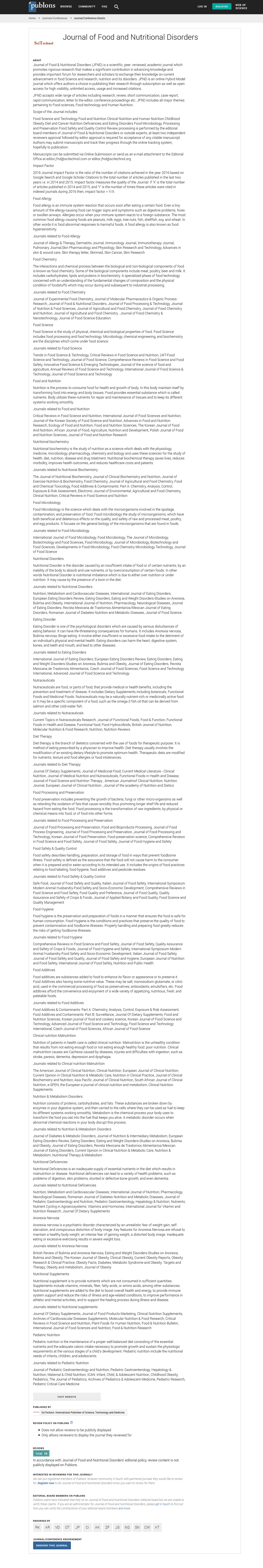
Induced type 1 diabetes mellitus caused an increase in the basophils, monocytes, and neutrophils while a significant decrease in the percentage of lymphocytes. The inducement did not cause any significant differences in the number eosinophils as reflected in Figure 1. Figure 1 shows the results obtained for the white blood cell differential count from the experimental rabbits’ blood. The result showed that group 3 (Induced and not treated) recorded the highest counts of basophils, monocytes, and neutrophils while recording the least count of lymphocytes. Lymphocytes were however higher in groups 1, 2 and 4 respectively with a value of 37.33%. These same groups also recorded close values of 54.33% neutrophils. The group induced with diabetes and not treated at all recorded the highest neutrophil count of over 65% which far exceed the values recorded for all the other groups. It is equally important to note that the group induced and treated with drug had the highest eosinophil. The group induced and not given any treatment (group 3) also had the highest monocytes of over 12% compared to other treatments which were less than 10%. The monocyte count for the group treated with drug after inducement of diabetes was least which is less than 8%. Basophil was also higher the group induced and not treated while it was least in groups 1, 4 and 5 (control; induced and treated with garlic juice; and induced and treated with orange juice).

Figure 1: Results of the differential counts of white blood cells from experimental rabbits. Keys: Group 1: Control; Group 2: Induced and treated with drug; Group 3: Induced and not treated; Group 4: Induced and treated with garlic juice; Group 5: Induced and treated with orange juice; Group 6: Induced and treated with yoghurt; Group 7: Induced and treated with fermented beniseed liquor.
The effect of type 1 induced diabetes on the weight of the experimental rabbits showed that diabetes caused a drastic decrease in weight of the rabbits. In group 1 (control) the weight of the rabbits increased steadily for the twelve (12) weeks of the experiment. The weight increased from 0.52 ± 0.05 kg at the first week to 1.21 ± 0.03 kg by the twelfth week. Contrastingly, the weight of the rabbits in the group that was induced with type-1 diabetes mellitus showed an increase in weight within the first four (4) weeks of the experiment from 0.59 ± 0.05 kg to 0.75 ± 0.05 kg before reducing to as low as 0.48 ± 0.02 kg by the tenth week of the experiment with significant differences at p ≤ 0.05. By the seventh (7th) week of the experiment, there were no significant differences at p ≤ 0.05 in the weight of the rabbits in week one (1). However, by the twelfth week, there was a little increase to 0.53 ± 0.05 kg with significant differences at p ≤ 0.05. Comparatively, the group induced and treated with garlic extract showed the effectiveness of the extract in the fact that there were significant differences at p ≤ 0.05 in weight of the rabbits from the first week of the experiment through to the last week of the experiment.
The effect of the weight on group 4 (the group induced and treated with garlic juice) was the fastest treatment to regain its weight. Although, there were no significant differences at p ≤ 0.05 between week 1 and week 3 (which is the period of inducement of diabetes), there was significant differences at p ≤ 0.05 in the weight of the rabbits positively for the rest of the research with the weight of the rabbits increasing from 0.61 ± 0.03 kg to 1.14 ± 0.11 kg at the twelfth week of the experiment. Comparatively, the results obtained for groups 5, 6 and 7 did not record much weight recovery as in group 4. For instance, in group 5 (the group induced and treated with orange juice) there were no significant differences at p ≤ 0.05 in the weight of the experimental rabbits between weeks 1, 6 and 7. The significant differences at p ≤ 0.05 in the weight of the rabbits were only observed between weeks 8 and 12 from 0.65 ± 0.03 kg to 0.79 ± 0.07 kg as an increase. In that of group 6 (induced and treated with yogurt) the significant differences at p ≤ 0.05 in the weight of the rabbits between the time of the inducement and the termination of the research at the twelfth week were less than 10 g. In group 7 (induced and treated with fermented beniseed liquor), the same pattern of result as in group 4 was observed. However, the effect of the treatment on the increase in weight was not as effective as in that of group 4. The effect of the treatments on the weight of the rabbits is summarized in Table 4.
| Week | Group 1 | Group 2 | Group 3 | Group 4 | Group 5 | Group 6 | Group 7 |
|---|---|---|---|---|---|---|---|
| 1 | 0.52 ± 0.05a | 0.52 ± 0.06a | 0.59 ± 0.05 e | 0.60 ± 0.00a | 0.61 ± 0.05a | 0.67 ± 0.05a | 0.68 ± 0.04c |
| 2 | 0.56 ± 0.04b | 0.77 ± 0.09 c | 0.64 ± 0.06 f | 0.65 ± 0.05b | 0.69 ± 0.01d | 0.85 ± 0.05f | 0.71 ± 0.06d |
| 3 | 0.59 ± 0.13b | 0.85 ± 0.10 d | 0.76 ± 0.02 h | 0.61 ± 0.03a | 0.76 ± 0.02f | 0.89 ± 0.07g | 0.79 ± 0.07g |
| 4 | 0.65 ± 0.15c | 0.81 ± 0.09 c | 0.75 ± 0.05 h | 0.69 ± 0.07c | 0.74 ± 0.00e | 0.90 ± 0.02g | 0.74 ± 0.02e |
| 5 | 0.68 ± 0.00c | 0.72 ± 0.05b | 0.70 ± 0.00 g | 0.78 ± 0.04d | 0.70 ± 0.04d | 0.85 ± 0.02f | 0.70 ± 0.00d |
| 6 | 0.73 ± 0.09c | 0.73 ± 0.11 b | 0.62 ± 0.10 f | 0.82 ± 0.00e | 0.62 ± 0.01a | 0.81 ± 0.07d | 0.65 ± 0.09b |
| 7 | 0.81 ± 0.11d | 0.79 ± 0.07 c | 0.60 ± 0.10 e | 0.90 ± 0.05f | 0.62 ± 0.06a | 0.75 ± 0.03b | 0.62 ± 0.06a |
| 8 | 0.85 ± 0.10e | 0.81 ± 0.03 c | 0.55 ± 0.11 d | 0.96 ± 0.02g | 0.65 ± 0.03b | 0.76 ± 0.05b | 0.68 ± 0.04c |
| 9 | 0.92 ± 0.02e | 0.86 ± 0.08 d | 0.51 ± 0.07 b | 0.99 ± 0.03h | 0.67 ± 0.03c | 0.79 ± 0.03c | 0.76 ± 0.02f |
| 10 | 0.97 ± 0.03f | 0.89 ± 0.11 e | 0.48 ± 0.02 a | 1.04 ± 0.01i | 0.69 ± 0.07d | 0.80 ± 0.04c | 0.81 ± 0.03h |
| 11 | 1.09 ± 0.13f | 0.92 ± 0.02 e | 0.51 ± 0.09 b | 1.10 ± 0.03j | 0.75 ± 0.05f | 0.83 ± 0.07e | 0.85 ± 0.07i |
| 12 | 1.21 ± 0.03g | 0.96 ± 0.08 f | 0.53 ± 0.05 c | 1.14 ± 0.11k | 0.79 ± 0.07g | 0.85 ± 0.05f | 0.87 ± 0.12j |
Keys: Group 1: Control; Group 2: Induced and treated with drug; Group 3: Induced and not treated; Group 4: Induced and treated with garlic juice; Group 5: Induced and treated with orange juice; Group 6: Induced and treated with yoghurt; Group 7: Induced and treated with fermented beniseed liquor
Table 4: Effect of diabetes on the weight (kg) of rabbits.
The results of the histopathology of the pancreas of the experimental rabbits used in this assay showed different pathological features based on the treatments the rabbits were subjected to. The control group (group 1) showed presence of cracked anterior artifacts with visible active acinus and well-formed cell infiltrations with well-spaced interstitial cells of the pancreatic ducts (Plate 1). Group 2, on the other hand, showed entirely different pathology in which the pancreatic lobular cells are prominent and centrally located with visible connective septae. There is presence of cell infiltrations and very few inflammatory cells packed around the islet of Langerhans. Sides’ globular pancreatic cells are not well-formed with slight anterior washing away of globular cells leading to intercellular drainage of exocrine secretions (Plate 2).

Plate 1: Pancreas from group 1 (Control) experimental rabbits.

Plate 2: Pancreas from group 2 (Induced and treated with drug) experimental rabbits.
The effects of the severely diabetic pancreas are well pronounced in the 3rd group of rabbits (group induced and not treated). The pathological features seen include poor formation of the islet of Langerhans cells and dot necrotized cells. There is profuse hemorrhage from highly vascularized pancreatic acini surrounded by parenchyma fat cells. Few intralobular ducts are seen without any visible interlobular duct on the plate (Plate 3). The group induced with diabetes and treated with garlic extract (Plate 4) showed some good features that not only depict a good and fast recovery from diabetes but showed but some other health benefits of garlic. They are presence of well-formed pancreatic acini and cell infiltrations with well-spaced interstitial cells of the pancreas that shows active cell division of the pancreatic ducts and acini. There is splay and intrafaradization of the cracked pancreatic ducts. The presence of artifacts is seen at the anterior portion of the plate with well-formed interlobular and intralobular ducts.

Plate 3: Pancreas from group 3 (Induced and not treated) experimental rabbits.

Plate 4: Pancreas from group 4 (Induced and treated with garlic juice) experimental rabbits.
The histopathology of the group induced and treated with orange juice (group 5), there is the poor formation of the islet of Langerhans cells with dot necrotized cells. There is the presence of profuse hemorrhage from highly vascularized pancreatic acini that is surrounded by parenchyma fat cells. Large dilated vacuoles of interlobular ducts with dot necrosis are seen scattered within the plate. The pancreatic cells of the group induced and treated with yogurt (group 6) have anlage pancreatic ducts lining the acinar lobules. There is presence of inflammatory cell infiltrations with their mesenteric intercalated ducts that are well-formed as well as proper intralobular ducts. The plate showed no visible interlobular ducts or hemorrhage but shows some anterior dot necrosis. The distal end acinar are not well-formed. The pancreatic lobular cells are prominent and centrally located with visible connective septae. There is presence of inflammatory cell infiltrations and sides’ globular pancreatic cells are not well-formed with slight anterior dot necrosis of globular cells leading to intercalated karyolysis of exocrine secretory duct. There is little posterior end hemorrhage. The results of the histopathology are shown in Plates 1-7.

Plate 5: Pancreas from group 5 (Induced and treated with orange juice) experimental rabbits.

Plate 6: Pancreas from group 6 (Induced and treated with yoghurt) experimental rabbits.

Plate 7: Pancreas from group 7 (Induced and treated with fermented beniseed liquor) experimental rabbits.
There is the presence of well-formed cell infiltrations with wellspaced interstitial cells of the pancreas that shows active cell division of the pancreatic ducts. There is splay and intrafaradization of the cracked pancreatic ducts, the presence of artifacts are seen at the anterior portion of the plate.
The pancreatic lobular cells are prominent and centrally located with visible connective septae. There is presence of cell infiltrations and very few inflammatory cells packed around the islet of Langerhans. Sides’ globular pancreatic cells are not well-formed with slight anterior washing away of globular cells leading to intercellular drainage of exocrine secretions.
There is the poor formation of the islet of Langerhans cells and dot necrotized cells. There is profuse hemorrhage from highly vascularized pancreatic acini surrounded by parenchyma fat cells. Few intralobular ducts are seen without any visible interlobular duct on the plate.
There is the presence of well-formed pancreatic acini and cell infiltrations with well-spaced interstitial cells of the pancreas that shows active cell division of the pancreatic ducts and acini. There is splay and intrafaradization of the cracked pancreatic ducts, the presence of artifacts are seen at the anterior portion of the plate. Wellformed interlobular and intralobular ducts.
There is poor formation of the islet of Langerhans cells and dot necrotized cells. There is profuse hemorrhage from highly vascularized pancreatic acini surrounded by parenchyma fat cells. There are large dilated vacuoles of interlobular ducts with dot necrosis scattered within the plate.
Pancreatic cells have anlage pancreatic ducts that lined the acinar lobules. There is the presence of inflammatory cell infiltrations. The mesenteric intercalated ducts are well-formed as well as the intralobular ducts. There are no visible interlobular ducts or hemorrhage but shows some anterior dot necrosis.
The distal end acinar is not well-formed. The pancreatic lobular cells are prominent and centrally located with visible connective septae. There is presence of inflammatory cell infiltrations and sides’ globular pancreatic cells are not well-formed with slight anterior dot necrosis of globular cells leading to intercalated karyolysis of the exocrine secretory duct. There is little posterior end hemorrhage.
Discussion
The inducement of type-1 diabetes on the rabbits using streptozotocin has pointed to the fact that streptozotocin can cause chronic diabetes especially when administered with a booster dose in rabbits. The shooting up of the sugar level from 70.02 ± 1.0 mg/ dl to as high as 187.33 ± 0.9 mg/dl after the third booster was given to the experimental rabbits has shown the chronic effect of the type 1 diabetes caused by streptozotocin. This result is similar to the results obtained in which streptozocin induced type 1 diabetes in rabbits increased the sugar level by 70%.
Equally, the effects of the induced type-1 diabetes on the gross morphology of the rabbits’ organs (liver, kidneys, and pancreas) are swellings or oedema consequently causing an increase in the weight of these organs when compared to the control. In a research conducted by Lei et al. [12] on rats’ organs using streptozotocin, it was concluded that diabetes can cause some level of morphological distortion of vital organs including swelling or oedema. From Table 2, only two groups showed “all normal” for the organs (The control group and the group induced and treated with garlic juice). This result, therefore, shows that diabetes can cause pancreas enlargement and even discoloration of liver hepatocytes.
The results of the PCV for the various treatments revealed the effects of the drug (Glucovance), garlic marsh, yogurt, orange juice, and fermented beniseed liqour not just on the blood sugar but also on the body system of the experimental rabbits used in this research. The fact that diabetes drug (glucovance) was able to lower the sugar level significantly different at p ≤ 0.05 is indicative of the effectiveness of the drug on diabetes. The results obtained for the PCV of the rabbits induced and treated with garlic extract which had the highest percentage is an insight to the blood-boosting nature ant therapeutic effect of garlic. In an experiment done to prove the therapeutic effect of garlic on rats induced with diarrhea by Adebolu et al. [13], garlic was able to stop the induced diarrhea as well as cause an increase in the blood level of the rats. Therefore, the results obtained for the PCV of the group induced and treated with garlic is a similar one. According to Toluwani [14], milk is a good blood enhancer and probiotics in fermented milk and milk products are aid digestion. This probably may be the reason why yogurt may be responsible for the secondhighest PCV values obtained.
Another point of significance of the RBC result. The result of the Red Blood Cell Count (RBC) was not significantly different at p ≤ 0.05 in the value obtained for groups 1, 2, 5 and 6 (control, induced and treated with drug, induced and treated with orange juice and induced and treated with yogurt) respectively. The group induced and treated with garlic was however significantly different at p ≤ 0.05 having the highest RBC value of 16.28 ± 1.54 when compared with the group induced and not treated that had a value of 9.02 ± 1.05. This result is similar to the result obtained by Latifat [15] in which the rats that were fed with yogurt and garlic extract had a very high RBC value than the control. The fibrinogen values obtained can best be described as high for other groups while it is normal for the control group and the group treated with garlic extract. Therefore, diabetes has negative effect on fibrinogen level of blood. This result corresponds with the result obtained by Madan et al. [16] in which diabetes increased the fibrinogen level of the blood samples examined. According to Kafle and Shrestha [17], this is usually the cause of hemorrhage or excessive bleeding in diabetic patients that result from poor clotting factors.
The WBC differential count results equally revealed the fact that type 1 diabetes mellitus caused an increase in the basophils, monocytes, and neutrophils while a significant decrease in the percentage of lymphocytes significantly. In related research by Vozarova et al. [18], high white blood cell count was found to be associated with a worsening of insulin sensitivity in rabbits. However, the group treated with garlic extract maintained the same level of all the differential counts with the control group. According to Oyetayo [19], the ability of garlic (when not taking in excess) to maintain a normal WBC differential value in animals makes it nutraceutical in nature.
The effect of diabetes on the weight of the animal cannot be overemphasized in this research. The drastic reduction of the weight of the experimental rabbits as a result of the diabetes-induced supports the fact that diabetes causes a reduction in weight of animals. Several authors have reported decrease in weight in related research involving rats and rabbits [15,16,18]. Equally, recovery and increase in weight were observed more for then group treated with garlic extract and drug. The islets of Langerhans play a critical role in glucose homeostasis. Islets are predominantly composed of insulin-secreting beta cells and glucagon-secreting alpha cells. In type 1 diabetes, the beta cells are destroyed by autoimmune destruction of insulin-producing beta cells resulting in hyperglycemia. This is a gradual process, taking from several months to decades. Much of the beta cell destruction takes place during a silent, asymptomatic phase. Therefore, the ability of garlic extract to increase the weight of the rabbits may be that it had the ability to stop the autoimmune destruction of insulin-producing beta cells.
The histopathology of the pancreas of the experimental rabbits used in this assay showed different pathological features based on the treatments the rabbits were subjected to and the effect of the healing the treatments were able to exert. The presence of cracked anterior artifacts with visible active acinus and well-formed cell infiltrations with spaced interstitial cells of the pancreatic ducts on the control is an indication of well-formed islet cells of the pancreas (Plate 1). According to Cheville [20], these signs are not negative pathology of the pancreas. They are rather signs that show that the pancreas is actively reproducing the islet cells. The features seen in group 2, on the other hand, are entirely different pathology in which the pancreatic lobular cells are prominent and centrally located with visible connective septae. The cell infiltrations and very few inflammatory cells packed around the islet of Langerhans are pathology of damages done to islet of Langerhans before a healing process indicated by the later end of the interpretation were initiated. Sides’ globular pancreatic cells that are not well-formed with slight anterior washing away of globular cells leading to intercellular drainage of exocrine secretions are healing process of injured pancreatic cells.
The effects of severely diabetic pancreas are well pronounced in the group of rabbits induced without administration of any treatment (group induced and not treated) showed negative pathological features that include poor formation of the islet of Langerhans cells and dot necrotized cells. The profuse hemorrhage from highly vascularized pancreatic acini surrounded by parenchyma fat cells showed that diabetes causes massive destruction islet cells [21]. Few intralobular ducts that are seen without any visible interlobular duct on the plate is an indication complete destruction of the islet cells better described as beyond repair. According to Gvazava et al. [22], once the pancreas of animals gets this bad, they mostly do not survive any form of treatment or therapy given to them in such state. The group induced with diabetes and treated with garlic extract (Plate 4) showed good features that not only depicts a good and fast recovery from diabetes but showed but some other health benefits of garlic. These include presence of well-formed pancreatic acini and cell infiltrations with well-spaced interstitial cells of the pancreas that shows active cell division of the pancreatic ducts and acini [22]. There is splay and intrafaradization of the cracked pancreatic ducts. The presence of artifacts is seen at the anterior portion of the plate with well-formed interlobular and intralobular ducts.
The histopathology of the group induced and treated with orange juice (group 5) which showed poor formation of the islet of Langerhans cells with dot necrotized cells clearly reveals the effect of the juice on diabetes. Although there was reduction in the sugar level of the blood as the experimental animals were being treated, it is however certain that orange juice alone as fruit cannot lower the blood sugar of a diabetic patient. According to Gvazava et al. [22], poor formation of islet of Langerhans accompanied with little necrosis is dangerous pathologically and calls for concern in therapeutic administration of any chemotherapy. These features were also similar to the features observed in group 6 in which pancreatic cells have anlage pancreatic ducts that lined the acinar lobules. The presence of inflammatory cell infiltrations and the mesenteric intercalated ducts that are well-formed, as well as the proper formation of intralobular ducts, are good signs of reproductive cells of the pancreas. However, the anterior dot necrosis in the group induced and treated with yogurt calls for concern.
The distal end acinars that are not well-formed and the pancreatic lobular cells that are prominent and centrally located with visible connective septae is a malformed tissue with the pathology of diseased state of the organ [23]. There is presence of inflammatory cell infiltrations and sides’ globular pancreatic cells that are not wellformed with slight anterior dot necrosis of globular cells leading to intercalated karyolysis of exocrine secretory duct account for the hemorrhage capable posing death threat to the rabbits. Gvazava et al. [22] listed these signs among the most critical signs of diabetic pancreas that will eventually cause the total breakdown of the entire system if not regulated on time.
The islets of Langerhans play a critical role in glucose homeostasis. Islets are predominantly composed of insulin-secreting beta cells and glucagon-secreting alpha cells. In type 1 diabetes, the beta cells are destroyed by autoimmune destruction of insulin-producing beta cells resulting in hyperglycemia [10]. This may be a gradual process, taking months or may be immediate when induced like the one in this research.
The effect of diabetes in the world today is alarming and calls for immediate research into nutraceutical foods that will aid the adequate recovery of patients without the use of the drug. The side effects of diabetic drugs could easily be prevented when these foods are used to treat diabetic patients. Also, these foods when taken constantly are capable of stabilizing the sugar level of the body. The effect of garlic extract as a good antidiabetic agent has been well established in this research. The curative strength of other foods such as yogurt and fruit extract has been proven in the findings of this research. Therefore, garlic has the most outstanding positive effect on diabetes and is recommended for people who are diabetic.
Funding Statement
This research was funded by Dr. (Mrs.) F. O. Omoya and Dr. A. O. Momoh as part of our research to meet up our employers’ demand for research, teaching and community service. Both authors (funders) were involved in the manuscript writing, editing, approval, and decision to publish this article accordingly.
Acknowledgments
Authors wish to acknowledge the following: the medical laboratory scientists of Adekunle Ajasin University, Akumgba; Staff of Obafemi Awolowo University Teaching Hospital, Ife; Veterinary section of Animal production and health of the Federal University of Technology, Akure and Staff of Biological Sciences Department of Elizade University, Ilara-Mokin. Thank you for your support.
Data Statement
The (Statistical Package for Social Sciences- SPSS version 22) data used to support the findings of this study are available from the corresponding author upon request.
Conflict of Interest
The authors hereby declare that there is no conflict of interest in this research.
References
- Doha AM, Minar MH (2017) Amelioration of diabetes in a rat model through yoghurt supplemented with probiotics and olive pomace extract. J Biol Sci 17: 320-333.
- Graham ML, Janecek JL, Kittredge JA, Hering BJ, Schuurman HJ (2011) The streptozotocin-induced diabetic nude mouse model: differences between animals from different sources. Comp Med 61: 356-360.
- Kitabchi AE, Umpierrez GE, Miles JM, Fisher JN (2009) Hyperglycaemic crises in adult patients with diabetes. Diabetes Care 32: 1335-13343.
- Adebolu TT, Adeoye OO, Oyetayo VO (2011) Effect of garlic (Allium sativum) on Salmonella typhi infection, gastrointestinal flora and hematological parameters of albino rats. Afr J Biotechnol 10: 6804-6808.
- Salih BA, Abasiyanik MF, Bayyurt N, Sander E (2007) H. pylori infection and other risk factors associated with peptic ulcers in Turkish patients: A retrospective study. World J Gastroenterol 13: 3245-3248.
- Srinivasan K, Ramarao P (2007) Animal model in type 2 diabetes research: An overview. Indian J Med Res 125: 451-472.
- Momoh AO, Adebolu TT, Ogundare AO (2012) Toxicological evaluation of fermented liquor and methanolic extract of beniseeds in albino rats. Int J Biopharm Nanomedical Sci 1: 58-63.
- Cheesbrough M (2006) District laboratory practice in tropical countries. Cambridge University Press.
- Baker FJ, Breach MR, Chris P (2014) Modern procedures for medical laboratory science. Chris Publisher, London, United Kingdom.
- Zotta E, Lago N, Ochoa F, Repetto HA, Ibarra C (2008) Development of an experimental hemolytic uremic syndrome in rats. Pediatr Nephrol 23: 559-567.
- Pagana KT (2017) Recent diagnostic, laboratory and histopathological test procedures, 4th Edition. St. Louis: Morsby.
- Lei YC, Hwang JS, Chan CC, Lee CT, Cheng TJ (2005) Enhanced oxidative stress and endothelial dysfunction in streptozotocin-diabetic rats exposed to fine particles. Environ Res 99: 335-343.
- Adebolu TT, Osoba EA, Momoh AO (2016) Comparative effect of oral rehydration therapy on the gastrointestinal flora and haematological parameters of albino rats. Afr J Biotechnol16: 7824-7831.
- Toluwani AO (2017) Assessment of probiotics in fermenting milk and milk product. Undergraduate thesis, Elizade University, Ilara-Mokin, Nigeria.
- Latifat O (2018) The effect of yogurt on ulcer-induced and diabetes-induced rats. Undergraduate thesis, Elizade University, Ilara-Mokin, Nigeria.
- Madan R, Gupta B, Saluja S, Kansra UC, Tripathi BK, et al. (2010) Coagulation profile in diabetes and its association with diabetic microvascular complications. J Assoc Physicians India 58: 481-484.
- Kafle DR, Shrestha P (2010) Study of fibrinogen in patients with diabetes mellitus. Nepal Med Coll J 12: 34-37.
- Vozarova B, Weyer C, Lindsay RS, Pratley RE, Bogardus C, et al. (2002) High white blood cell count is associated with a worsening of insulin sensitivity and predicts the development of type 2 diabetes. Diabetes 51: 455-461.
- Oyetayo VO (2014) Functional and nutraceutical foods. University of Ibadan press.
- Cheville NF (2009) Ultrastructural Pathology. Willey-Blackwell.
- Brezniceanu ML, Liu F, Wei CC, Chénier I, Godin N, et al. (2008) Attenuation of interstitial fibrosis and tubular apoptosis in db/db transgenic mice overexpressing catalase in renal proximal tubular cells. Diabetes 57: 451-459.
- Gvazava IG, Rogovaya OS, Borisov MA, Vorotelyak EA, Vasiliev AV (2018) Pathogenesis of type 1 diabetes mellitus and rodent experimental models. Acta Naturae 10: 24-33.
- Elsner M, Guldbakke B, Tiedge M, Munday R, Lenzen S (2000) Relative importance of transport and alkylation for pancreatic beta-cell toxicity of streptozotocin. Diabetologia 43: 1528-1533.