Research Article, Lpmj Vol: 106 Issue: 4
Serum response of cortisol and Troponin-I to endurance exercise and electrical stimulation in myocardial infarction rats
Mohammad Malekipooya1 Department of Sports Sciences, Faculty of Literature and Humanities, Islamic Azad University, Professor Hasabi Branch, Tafresh, Iran.
2 Department of Sports Sciences, Faculty of Literature and Humanities, Islamic Azad University of Arak, Arak, Iran.
3 Department of Physiology, Faculty of Basic Sciences, Arak University of Medical Sciences, Arak, Iran.
4 Department of Sports Sciences , Faculty of Sports Sciences, Arak University, Arak, Iran. 5-Department of Sports Sciences, Islamic Azad University, Mahallat Branch, Mahallat, Iran.
*Corresponding Author : Mohammad MalekiPooya Department of Sport Sciences, Faculty of Literature and Humanities E-mail: Malekipooya@iautb.ac.ir Tel: +98-086-6238502
Received Date: July 02, 2020; Accepted Date:July 11, 2020; Published Date: July 29, 2020
Citation: Mimbale J (2020) Pre-operative predictors of difficult laparoscopic cholecystectomy. LPMJ 106:4.
Abstract
Background and Aim: Due to the high prevalence of myocardial infarction and an increase in deaths caused by it by 2030, it is essential to provide alternative therapies. The aim of the present study was to investigate the serum response of CORT and cTnI to endurance exercise and electrical stimulation in myocardial infarction rats.
Materials and Methods: This experimental study was conducted with post-test design and with a control group. After induction of infarction with two subcutaneous injections of isoproterenol (150 mg / kg), 50 Wistar rats (8 weeks with a weight of 230 ± 30 g) were randomly divided into 5 groups of healthy, infarction, infarction-endurance exercise, infarction-electrical stimulation, and infraction-endurance exercise-electrical stimulation. The intervention groups received electrical stimulation for one session (foot shock device with a current intensity of 0.5 mA and a duration of 20 min) and endurance exercise (treadmill at a speed of 20 m / min for 1 h). Immediately after the intervention, serum levels of CORT and cTnI were measured. ANOVA and Tukey tests were used to analyze the data at a significance level of P <0.05.
Results: The results showed that myocardial infarction significantly increased serum levels of CORT and cTnI (P <0.0001). Also, the concentration of CORT in endurance exercise groups (P = 0.0008), electrical stimulation (P = 0.032) and endurance exercise-electrical stimulation (P = 0.044) showed a significant reduction compared to myocardial infarction group. Also, cTnI concentration in endurance exercise group showed a significant reduction compared to myocardial infarction group (P = 0.013).
Conclusion: It seems that endurance exercise and electrical stimulation individually and in combination with each other can result in an improvement in the conditions of myocardial infarction patients by reducing the concentration of CORT and cTnI
Keywords: Myocardial Infarction, Electrical Stimulation, Troponin-I, Endurance Exercise, CORT
Introduction
Cardiovascular disease is one of the leading causes of death in the world [1] and by 2030 it will account for more than 30.5% of all deaths, of which 80% occur in low- and middle- income countries [2]. Ischemia myocarditis is caused by obstruction of the coronary arteries and manifests with clinical conditions such as angina pectoris, irregular heartbeats, heart failure, myocardial infarction (MI), or sudden death. It leads to the death of myocyte cells due to loss of blood flow, anemia, and ischemia [3]. Also, MI is one of the most common causes of coronary heart disease [4] , which by reducing and stopping blood flow, it has drawn the attention of cardiologists more than any other factor involved in heart attack. MI-induced necrosis stimulates the hypothalamic-pituitary-adrenal axis and, as a stressor, increases cortisol (CORT) and catecholamine levels [5, 6]. High levels of CORT increase mortality in MI patients [7-9]. Thus, the most appropriate method to cope with this disease is to identify the main risk factors and try to moderate them. Several studies have shown a link between stress and cardiovascular disease. Stress increases the secretion of catecholamines and corticosteroids from the endocrine glands, and subsequently, high levels of these hormones potentially increase the risk of cardiovascular disease [10]. The American Heart Association has also shown that stress reduces myocardial tissue blood flow by increasing the need for oxygen, increasing vascular resistance, and contracting coronary artery and it is considered a risk factor for cardiovascular disease [11].
Also, stress plays a major role in increasing CORT levels and this hormone has a long-term function against stress [12]. In a study conducted by Rao et al, results showed a direct link between CORT concentration and mortality in MI patients [13]. Studies have shown that exercise has been an effective stimulant on the hypothalamicpituitary- adrenal axis [14] and has led to an increase in the secretion of adrenocorticotropin from the pituitary gland, which is the most important factor in CORT secretion. The results of some other studies also show a significant reduction in CORT levels [15]. Evidence also suggests that there is a direct association between CORT and Troponin-I (cTnI) levels in MI patients. These factors are secreted due to the hypoxia-induced compatibility [5]. Troponin is one of the most sensitive proteins in MI damage conditions. The troponin complex consists of subunits of I, T, and C, which are attached to the thin filaments of myofibrils. Along with calcium, this protein plays a crucial role in regulating muscle contraction. Separate cTnI and cTnT isoforms are present in cardiac myocytes and, when released into the blood, it can be specifically measured by immunoassay methods [16]. cTnI and cTnT are also biomarkers of myocardial damage and are a standard factor in the diagnosis of MI caused by cardiac ischemia [17]. Due to having 31 amino acids in its N-terminal, cTnI is more specific than the other two components, which these amino acids are not found in other isomers. cTnI levels increase immediately after myocardial damage and return to normal level within 7 days [18]. Studies have shown that long-term continuous exercise can create cTnI [19]. Studies have shown that exercise intensity and duration are important factors in increasing cardiac troponin levels [20]. In this regard, Shaw et al reported that cTnI is released in response to intense short-term exercise [21]. Also, Vakshleg et al did not observe significant changes after intense short-term exercise on cTnI of dogs [22]. Also, in another study, no significant changes were reported following high-intense and continuous intermittent exercise in cTnI of healthy young adults [23]. In the study conducted by Karanza Garcia et al, intermittent activities did not affect the serum concentration of cTnI in healthy men [24]. The results of the studies show that sports activities cause different responses at CORT and cTnI levels. Different treatments have been used in MI patients. One of these methods, along with exercise, is the use of electrical stimulation (ES) in the rehabilitation of these patients. Based on the studies conducted, ES is expected to be a rehabilitation method for people who participate in exercise [25].
ES has also been used as a new and effective modality in the treatment of ischemia [26]. Since the effect of endurance exercise and electrical stimulation on CORT and cTnI changes in infarction samples has not been investigated so far, the present study was conducted to investigate the Cortisol and Troponin-I serum response to endurance exercise and electrical stimulation in MI rats.
Materials and Methods
Samples and research environment
This applied study was conducted by experimental design by posttest method and with a control group. In the present study, a total of fifty 8-week-old Wistar male rats with a mean weight of 230± 30 were used. Samples were transferred to the laboratory for one week to adapt to the environment and stored in transparent polycarbonate cages and under controlled environmental conditions with a temperature of 22 ±2 ° C, a humidity of 50±5 and a light-dark cycle of 12:12 hours with a free access to water and food. Then, they were randomly divided into 5 groups of healthy ones (H), myocardial infarction (MI), myocardial infarction-endurance exercise (MI.EX), myocardial infarctionelectrical stimulation (MI.ES) and myocardial infarction-endurance exercise- electrical stimulation (MI.EX.ES) (each group included 10 subjects).
Induction of myocardial infarction
Subcutaneous injection of isoproterenol (manufactured by Sigma USA) at a dose of 150 mg per kg body weight was used to induce MI [27]. Isoproterenol diluted with normal saline was injected subcutaneously for two consecutive days at 24-hour intervals. It is one of the most common methods of inducing MI in animal models, especially rats [28]. After 48 hours of the last injection, a number of rats were randomly selected from each group and underwent experimental conditions to ensure induction of infarction. In this study, MI was confirmed based on electrocardiographic changes (elevated ST segment) along with increased cardiac enzyme of cTnI (344.01 pg / ml).
Electrical stimulation program
To create the electrical stimulation protocol, the R12Electromodule Stimulator device manufactured by Partow Danesh Company was used. In this protocol, the current intensity of 0.5 mA was considered for 20 minutes. It was sent to the foot shock device through the output of the stimulator with the settings of Trial Number: 1, Trial Period: 1200000, Recording Time: 1200000.
Endurance exercise program
The rats were familiarized with the treadmill for 1 week, 5 days per week, 10 minutes each day at a speed of 10 m / min [29]. Investigations have shown that this level of exercise is not enough to cause a significant change in aerobic capacity. The rats were conditioned via sound and stimulation to avoid approaching, resting, and colliding with the electric shock
part at the end of the treadmill. Endurance exercise program was performed as one session of running on the treadmill for 1 hour, at speed of 20 m / min, without incline and about 60% of the maximal oxygen consumption [30].
Blood sampling and biochemical evaluation
The groups were anesthetized and killed immediately after the end of the exercise protocol with a combination of ketamine (75 mg / kg) and xylazine (10 mg / kg). At various stages, moral issues were observed and it was tried to avoid any physical harm and unnecessary methods. After the anesthesia, blood sampling was performed directly from the right atrium of the rats’ heart with 10-cc syringes. The taken blood was poured into simple chelated gel- containing tubes and, after being kept at room temperature for 10 minutes and clotting, it was centrifuged for 5 minutes at 5000 rpm. Then, serum samples were placed at a temperature of - 80 ° C for biochemical analysis. To determine the serum values of CORT and cTnI, ELISA method was used according to the instructions of the kits manufactured by Company of Eastbiopharm (with the intra-assay coefficient of variations of less than 12% for both kits and sensitivity of the measurement method of 0.52 ng / ml and 2.52 pg/ml, respectively).
Statistical Analysis
After confirming the normal distribution of data using the Shapiro- Wilk test, a one-way analysis of variance (ANOVA) and Tukey post-hoc tests were used to compare the means between the groups. Statistical analysis was performed using GraphPad (Version 6) software at the significance level (P <0.05) and 95% confidence level.
Ethical considerations
The present study was approved with Code of Ethics of IR.IAU.ARAK. REC.1397.007 by the Research Ethics Committee of the Islamic Azad University, Arak Branch.
Results
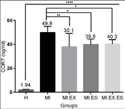
Chart 1 shows the serum concentration of CORT in the studied groups after the intervention. The results showed that serum CORT levels in rats had a statistically significant difference between healthy group and all groups (F=15.1 and P=0.0001). Also, CORT levels have a statistically significant difference between MI and MI.EX groups (F=5.2 and P=0.008), between MI and MI.ES (F=4.4 and P=0.032) and between MI and MI.EX.ES (F=4.2 and P=0.044). Also, Tukey test did not show a statistically significant difference between MI.EX and MI.ES (F=0.8 and P=0.980) and between MI.ES and MI.EX.ES (F=0.1
Chart 1: Changes in serum concentrations of CORT in healthy group: H, Myocardial infarction group: MI, endurance exercise + myocardial infraction group: EX.MI, myocardial infraction + endurance exercise + electrical stimulation group: MI.EX.ES show significant differences at the levels of (*P <0.05 (*P <0.01) and (*P <0.0001)
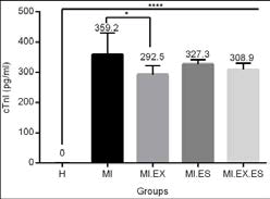
Chart 2 shows the serum concentration of cTnI in the studied groups after the intervention. The results showed that the levels of cTnI in healthy rats had a statistically significant difference with all groups (F=26.3 and P=0.0001). Also, the cTnI levels showed a significant difference between the MI and MI.EX groups (F=4.9 and P=0.013). Also, the difference between the MI and MI.ES (F=2.3 and P=0.476) and MI.EX.ES groups (F=3.7 and P=0.094) was not significant. Also, the Tukey post-hoc test did not show significantly a difference between the MI.EX and MI.ES groups (P=2.5 and P=0.390) and between MI.ES and MI.ES groups (F=1.2 and P=0.911), and between MI.ES and MI.EX.ES groups (F=1.3 and P=0.873).
Chart 2: Changes in serum concentrations of cTnI in healthy group: H, Myocardial infarction group: MI, endurance exercise + myocardial infraction group: EX.MI, myocardial infraction + endurance exercise + electrical stimulation group: MI.EX.ES, and significant difference at the levels of (*P<0.05 (*P<0.01) and (*P<0.0001).
Discussion
The present study was conducted to investigate changes in serum CORT and cTnI values after endurance exercise and electrical stimulation in MI samples. The results revealed that induction of MI significantly increased serum levels of CORT and cTnI. The results of the present study suggest that one session of endurance exercise significantly reduces CORT and cTnI- levels in MI samples. Cortisol is one of the most important steroid hormones that is secreted into the blood flow in cases of severe stress and is effective in regulating cardiovascular, immunological, homeostasis, and metabolic functions. It also accelerates the gluconeogenesis, lipogenesis, and proteolysis in body [31].
In a study in line with the present study, Klapersky et al showed a decrease in CORT values after endurance exercise [32]. Also, in another two studies, a reduction in CORT values were reported following endurance and strength exercises [33, 34]. In contrast to results of the present study, Zu et al showed that acute aerobic exercise increases CORT levels in both males and females [35]. In another inconsistent study, results showed that acute interval and intermittent exercises increased CORT values in rats [36]. The inconsistency in results of the present study and those of mentioned studies can be attributed to differences in the intensity of physical activities. In the present study, the intensity of the exercise was about 55% vo2max (moderate intensity) and it has been already shown that moderate and low intensity physical activities do not cause significant changes in CORT level, but high intensity physical activities stimulate the pituitary-hypothalamic axis, leading to an increase in CORT [37, 38].
The present study revealed a significant reduction in cTnI levels after one session of endurance exercise in myocardial infraction rats. In line with the results of the present study, Marefati et al showed that interval exercise with moderate intensity significantly reduced cTnI serum levels in rats with ischemia, indicating a protective role of this type of exercise against ischemic injury [39]. Reconstruction of muscle fibers and connective tissue changes after an exercise program can be one of the reasons for the decrease in cTnI levels [40] . In contrast to results of the present study, Noano et al showed that exercise significantly increased cTnI values in rats with ischemic MI [41]. In another study, one session of long-term exercise with high intensity significantly increased cTnI-levels in rats [42]. In this regard, Alsoyan examined the effect of short and long- term exercise on cardiac changes and showed a significant increase in cTnI in the short-term exercise group [43]. Since studies have shown that in addition to the intensity and duration of exercise, other important factors such as ambient temperature, humidity, altitude, dehydration, gender, and age may be effective in determining the onset of exercise-induced fatigue and heart damage, inconsistency in the results can be attributed to the abovementioned factors [44, 45]. An increase in cTnI secretion has not been accurately clarified after long-term activity, but this increase may be due to oxidative stress, hypoxia, unstable secretion caused by cytosolic leakage due to ischemia, change in cell membrane permeability and necrosis of cardiac cells [46]. It has been also reported that CTnI serum concentration reach its peak three to six hours after exercise and return to baseline 24 hours later. Therefore, difference in time of measuring cTnI levels can be another reason for inconsistency in the results of the mentioned studies and those of the present study [23]. Electrical stimulation (ES) is used as a new and effective modality in the treatment of ischemia [26], which was used as another intervention in this study. Results of the present study showed that the induction of ES in myocardial infraction rats significantly reduced CORT levels. In contrast to the present study, Datjet et al studied the acute and chronic effects of foot shock ES (0.8 mA and 20 min) on rats and showed that CORT and ACTH levels increased significantly [47]. In another inconsistent study, results showed that the acute foot shock ES (60 min, 1 mA, and 1 Hz) significantly increased CORT levels [48]. Also, the stressreducing effect of Furosemide in foot shock ES models in rats resulted in an increase in plasma CORT levels [49].
The discrepancy in the results can be due to differences in the intensity and duration of electrical stimulation induced on the study samples. In the mentioned inconsistent studies, the induced duration was more than 20 minutes and the electric current intensity was more than 0.5 mA. However, in the present study, the intensity was 0.5 mA and the duration of the electrical stimulation protocol was 20 minutes. The results of ES intervention on cTnI levels of myocardial infraction rats did not show a significant difference. In this study, it was shown that the combined effect of endurance exercise and ES significantly reduces the CORT levelin all study groups compared to MI. However, the cTnI changes in the endurance exercise plus ES group were not significant compared to the MI group. Researchers were not found a study to investigate the effect of foot shock ES on cTnI serum levels. Also, the researchers did not study to examine the effect of foot shock ES on cTnI serum levels and combination of endurance exercise and ES on cTnI and CORT serum values.
Conclusion
It seems that conducting one session of endurance exercise and electrical stimulation individually and in combination with each other leads to an improvement in the condition of MI patients by reducing the concentration of CORT and cTnI.
Authors contribution
All authors met all standard criteria of writing based on the recommendations of the International Committee of Medical Journal Publishers.
References
- Clark, H., NCDs: a challenge to sustainable human development. Lancet 2013(381): p. 510–1.
- Mozaffarian D, B.E., Go AS, Arnett DK, Blaha MJ, Cushman M, et al, Heart disease and stroke statistics--2015 update: a report from the American Heart Association. Circulation, 2015. 131(4): p. e29-322.
- Sugiyama A, H.Y., Okada M, Yamawaki H., Endostatin Stimulates Proliferation and Migration of Myofibroblasts Isolated from Myocardial Infarction Model Rats. International journal of molecular sciences., 2018. 3(19): p. 741.
- O’Gara PT, K.F., Ascheim DD, Casey DE, Chung MK, de Lemos J a, et al, ACCF/AHA guideline for the management of ST-elevation myocardial infarction: a report of the American College of Cardiology Foundation/American Heart Association Task Force on Practice Guidelines. Journal of American College of Cardiology 2013 4(61): p. e78- e140.
- Gann DS, F.A., Endocrinology and metabolic response to injury., ed. t. ed. 1994: New York: Mc Graw-Hill gb Inc.
- Vetter NJ, A.W., Strange RC, Oliver MF, Initial metabolic and hormonal response to acute myocardial infarction. Lancet, 1974. 1(7852): p. 284-8.
- Mayer, S., Effect of catecholamines on cardiac metabolism. Circ Res, 1974. 34(3): p. 129-135.
- Reis, D., Potentiation of the vasoconstrictor action of tropical norepinephrine on the human bulbar conjungtival vessel after tropical application of certain adrenocortical hormones. J Clin Endocrinol & Metab 1959. 20: p. 446-56.
- McAlpine HM, M.J., Leckie B, Rumley A, Gillen G, Dargie HJ, Neuroendocrine activation after acute myocardail infarction. Br Heart J 1988. 60: p. 117-24.
- Brydon L, E.S., Jia H, Mohamed-Ali V, Zachary I, Martin GF, Steptoe A., Psychological stress activates interleukin-1 beta gene expression in human mononuclear cells. . Brain Behav Immun., 2005. 19(6): p. 540-6.
- Curfinan, D., Is Exercise Beneficial: or Hazardous: To your Heart? NEJM 1993. 23: p. 1730-1731.
- Qureshi GM, S.G., Soomro AM, Pirzado ZA, Abbasi SA., Association of blood IL-6, Cortisol and Haemodynamics in Healthy adults with stress:A possible role in early atherogenesis. Sindh Univ Res J., 2009. 41(2): p. 41-6.
- Rao M.Sudhakar, D.T., Kareem Hashir, Padmakumar R, Ashwal A J. , Serum Cortisol Level as a Predictor of In-Hospital Mortality in Patients Undergoing Primary Percutaneous Intervention for ST Segment Elevation Myocardial Infarction. International Journal of Cardiovascular Practice, 2019. 4(1): p. s.
- Budde H, M.S., Ribeiro P, Wegner M. , The cortisol response to exercise in young adults. Front Behav Neurosci., 2015. 9(13).
- Ramanjaneya M, C.J., Brown JE, Tripathi G, Hallschmid M, Patel S, et al., Identification of nesfatin-1 in human and murine adipose tissue: a novel depot-specific adipokine with increased levels in obesity. Endocrinology 2010. 151(7): p. 3169-80.
- Apple FS, J.R., Newby LK, et al. , National Academy of Clinical Biochemistry and IFCC Committee for Standardization of Markers of Cardiac Damage Laboratory Medicine Practice Guidelines: analytical issues for biochemical markers of acute coronary syndromes. . Clin Chem. , 2007. 53(4): p. 547-51.
- Thygesen K, A.J., Jaffe AS, et al. , Writing Group on behalf of the Joint ESC/ACCF/AHA/WHF Task Force for the Universal Definition of Myocardial Infarction.Third Universal Definition of Myocardial Infarction. J Am Coll Cardiol., 2012. 60(16): p. 1581-98.
- Morrow DA, C.C., Jesse RL, et al., National Academy of Clinical Biochemistry. National Academy of Clinical Biochemistry Laboratory Medicine Practice Guidelines: clinical characteristics and utilization of biochemical markers in acute coronary syndromes. . Circulation, 2007. 53(13): p. 556-74.
- Rifai N, D.P., O’Toole M, Rimm E, Ginsburg GS. , Cardiac troponin T and I, electrocardiographic wall motion analyses, and ejection fractions in athletes participating in the Hawaii Ironman Triathlon. The American journal of cardiology. , 1999. 83(7): p. 1085-9.
- Shave R, B.A., George K, Wood M, Scharhag J, Whyte G ,et al. , Exercise induced cardiac troponin elevation: evidence, mechanisms, and implications. Journal of the American College of Cardiology. , 2010. 56(3): p. 169-76. .
- Shave R, R.P., Low D, George K, Gaze D. , Cardiac troponin I is released following high-intensity short-duration exercise in healthy humans. . International journal of cardiology., 2010. 145(2): p. 337-9.
- Wakshlag J, K.M., Gelzer A, Downey R, Vacchani P. , The Influence of HighIntensity Moderate Duration Exercise on Cardiac Troponin I and CReactive Protein in Sled Dogs.Journal of Veterinary Internal Medicine. , 2010. 24(6): p. 1388-9.
- ., N.G., The Effects of Sprint Interval vs. Continuous Endurance Training on Physiological And Metabolic Adaptations in Young Healthy Adults. . Journal of human kinetics. , 2014. 44(1): p. 97-109.
- Carranza-García L.E, G.K., Serrano-Ostáriz E, Casado-Arroyo R, Caballero-Navarro A.L, & Legaz-Arrese A., Cardiac biomarker response to intermittent exercise bouts. . International Journal of Sports Medicine. , 2011. 32(5): p. 327-31.
- Dobsk P, N.M., Siegelov J, et al, Low-frequency electrical stimulation increases muscle strength and improves blood supply in patients with chronic heart failure. . Circ J, 2006. 70(1): p. 75-82.
- Patterson, C.a.R., M. S., Therapeutic angiogenesis: the new electrophysiology?
- Bertinchant J, R.E., Polge A, Marty-Double C, Fabbro-Peray P, Poirey S, et al. , Comparison of the diagnostic value of cardiac troponin I and T determinations for detecting early myocardial damage and the relationship with histological findings after isoprenaline-induced cardiac injury in rats. Clin Chim Acta, 2000. 298((1-2).): p. 13-28.
- Lobo Filho HG, F.N., Sousa RBd, Carvalho ERd, Lobo PLD, Lobo Filho JG. , Experimental model of myocardial infarction induced by isoproterenol in rats. Brazilian Journal of Cardiovascular Surgery. , 2011. 26(3): p. 469-76.
- Uysal, N., Sisman, A. R., Dayi, A., Ozbal, S., Cetin, F., Baykara, B., et al., Acute footshock-stress increases spatial learning–memory and correlates to increased hippocampal BDNF and VEGF and cell numbers in adolescent male and female rats. Neuroscience Letters 2012. 514(2): p. 141– 146.
- Schefer V, T.M., Oxygen consumption in adult and AGED C57BL/6J mice during acute treadmill exercise of different intensity. Exp Gerontol, 1996. 31(3): p. 387-392.
- Loria P, O.S., Michelazzi L, Giuria R, Ghisellini P, et al., Salivary Cortisol in an Extreme Non- Competitive Sport Exercise. Winter Swimming. Natu Sci, 2014. 6: p. 387-398
- Klaperski S, v.D.B., Heinrichs M, Fuchs R. l., Effects of a 12-week endurance training program on the physiological response to psychosocial stress in men: a randomized controlled trial. J Behav Med, 2014. 37(6): p. 1118-33.
- Rosa, G., Dantas, Estélio HM, & Mello, DB, The response of serum leptin, cortisol and zinc concentrations to concurrent training. Hormones, 2011. 10(3): p. 216-222.
- Schumann, M., Walker, Simon, Izquierdo, Mikel, & et all, The order effect of combined endurance and strength loadings on force and hormone responses: effects of prolonged training. European journal of applied physiology, 2014. 114(867-880).
- Zhou W, Z.G., Lyu C, Kou F, Zhang S, Wei H., The Effect of Exhaustive Exercise on Plasma Metabolic Profiles of Male and Female Rats. Journal of Sports Science and Medicine., 2019. 18(2): p. 253-263.
- De Araujo GG, G.C., Marcos-Pereira M, Dos Reis IG, Verlengia R, Interval versus continuous training with identical workload: physiological and aerobic capacity adaptations. Physiol Res. , 2015. 64(2): p. 209-19.
- Deane Jacks, J.S., John Anning, Thomas Mcgloughlin, Fredrick Andres., Effect of Exercise at Three Exercise Intensities on Salivary Cortisol. Journal of Strength and Conditioning Research., 2002. 16(2): p. 286-289.
- Hellhammer DH, W.S., Kudielka BM, Salivary cortisol as a biomarker in stress research. Psychoneuroendocrinology, 2009. 34(2): p. 163-71.
- Marefati, H., Aminizadeh, S., Najafipour, H., Dabiri, Sh. and Shahouzehi, B., The Effects of Moderate-Intensity Interval Training on the Resistance to Induced Cardiac Ischemia in Adult Male Rats. . Qom University of Medical Science Journal, 2016. 10(4): p. 1-9.
- Mehta R, G.D., Mohan S, Williams K, Sprung V, George K, et al, Postexercise cardiac troponin release is related to exercise training history. International Journal of Sports Medicine., 2012. 33(5): p. 333-7.
- Nounou, H.A., Deif, M. M., & Shalaby, M. A. ). Effect of flaxseed supplementation and exercise training on lipid profile, oxidative stress and inflammation in rats with myocardial ischemia. Lipids in Health and Disease, 2012. 11(1): p. 129.
- Chen, Y., Serfass, R. C., Mackey-Bojack, S. M., Kelly, K. L., Titus, J. L., & Apple, F. S, Cardiac troponin T alterations in myocardium and serum of rats after stressful, prolonged intense exercise. Journal of Applied Physiology, 2000. 88(5): p. 1749–55.
- N.S, A., Effect of Exercise and Vitamin E on Cardiac Troponin Alterations in Myocardium and Serum of Rats after Stressful Intense Exercise. International Journal of Zoological Research, 2010. 6(1): p. 24-29.
- Middleton N, S.R., Is exercise-induced cardiac fatigue caused by damage to the heart muscle and peak performance? . 2007.
- P, L., The evaluation and application of cardiac damage markers. In Biochemistry and Biophysics, 2006.
- Sato Y, K.T., Takatsu Y, Kimura T, Biochemical markers of myocyte injury in heart failure. Heart 2004. 90(10): p. 1110-1.
- Dagyte G1, V.d.Z.E., Postema F, Luiten PG, Den Boer JA, Trentani A, Meerlo P., Chronic but not acute foot-shock stress leads to temporary suppression of cell proliferation in rat hippocampus. Neuroscience., 2009 162(4): p. 904-13.
- Yutaka Endo, K.Y., Yukiko Fueta, Masahiro Irie, Changes of body temperature and plasma corticosterone level in rats during psychological stress induced by the communication box. Med Sci Monit, 2001. 7(6): p. 1161-65.
- Kaur, A., Bali, A., Singh, N., & Jaggi, A. S, Investigating the stress attenuating potential of furosemide in immobilization and electric foot-shock stress models in mice. Naunyn- Schmiedeberg’s Archives of Pharmacology, 2015. 388(5): p. 497–50.
 Spanish
  Chinese
  Russian
  German
  French
  Japanese
  Portuguese
  Hindi
Hindi 


